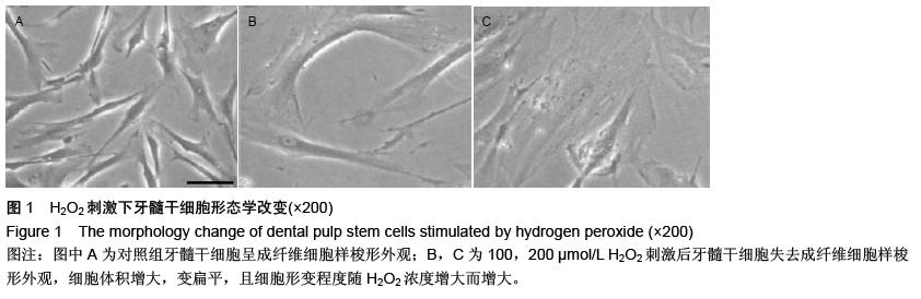
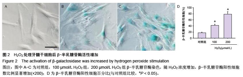
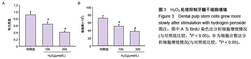
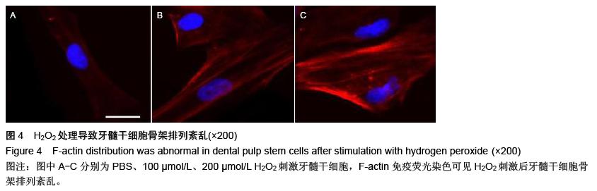
| [1] Gronthos S, Mankani M, Brahim J, et al. Postnatal human dental pulp stem cells (DPSCs) in vitro and in vivo. Proc Natl Acad Sci U S A. 2000;97(25): 13625-13630.[2] Lauritano D, Avantaggiato A, Candotto V, et al. Insulin activity on dental pulp stem cell differentiation: an vitro study.J Biol Regul Homeost Agents. 2015;29(3 Suppl 1):48-53.[3] Malekfar A, Valli KS, Kanafi MM, et al. Isolation and Characterization of Human Dental Pulp Stem Cells from Cryopreserved Pulp Tissues Obtained from Teeth with Irreversible Pulpitis. J Endod. 2016;42(1):76-81.[4] Laino G, d'Aquino R, Graziano A, et al. A new population of human adult dental pulp stem cells: a useful source of living autologous fibrous bone tissue (LAB). J Bone Miner Res. 2005;20(8):1394-1402.[5] Laino G, Carinci F, Graziano A, et al. In vitro bone production using stem cells derived from human dental pulp. J Craniofac Surg. 2006;17(3):511-515.[6] Arora V, Arora P, Munshi AK. Banking stem cells from human exfoliated deciduous teeth (SHED): saving for the future. J Clin Pediatr Dent. 2009;33(4):289-294.[7] Kolf CM, Song L, Helm J, et al. Nascent osteoblast matrix inhibits osteogenesis of human mesenchymal stem cells in vitro. Stem Cell Res Ther. 2015;6:258.[8] Hu X, Yang J, Wang Y, et al. Mesenchymal stem cells preconditioned with trimetazidine promote neovascularization of hearts under hypoxia/ reoxygenation injury. Int J Clin Exp Med. 2015;8(9): 16991-17005.[9] Qiao PF, Yao L, Zhang XC, et al. Heat shock pretreatment improves stem cell repair following ischemia-reperfusion injury via autophagy.World J Gastroenterol. 2015;21(45):12822-12834.[10] Morabito R, Romano O, La Spada G, et al. H2O2-Induced Oxidative Stress Affects SO4= Transport in Human Erythrocytes. PLoS One. 2016; 11(1):e0146485.[11] Zhou L, Chen X, Liu T, et al. Melatonin reverses H2 O2 -induced premature senescence in mesenchymal stem cells via the SIRT1-dependent pathway. J Pineal Res. 2015;59(2):190-205.[12] Shao J, Yang X, Liu T, et al. Autophagy induction by SIRT6 is involved in oxidative stress-induced neuronal damage. Protein Cell. 2016 Mar 16. [Epub ahead of print][13] 刘洋,吴晓冰,荆永光,等.氧化氢诱导间充质干细胞衰老模型的建立[J].军事医学, 2015,39(5):329-333.[14] Cheng H, Qiu L, Ma J, et al. Replicative senescence of human bone marrow and umbilical cord derived mesenchymal stem cells and their differentiation to adipocytes and osteoblasts Mol Biol Rep. 2011;38(8): 5161-5168.[15] Gu Z, Tan W, Feng G, et al. Wnt/β-catenin signaling mediates the senescence of bone marrow-mesenchymal stem cells from systemic lupus erythematosus patients through the p53/p21 pathway. Mol Cell Biochem. 2014;387(1-2):27-37.[16] Zhou D, Yin D, Xiao F, et al. Expressions of Senescence-Associated β-Galactosidase and Senescence Marker Protein-30 are Associated with Lens Epithelial Cell Apoptosis. Med Sci Monit. 2015;21: 3728-3735.[17] Gu Z, Tan W, Feng G, et al. Wnt/β-catenin signaling mediates the senescence of bone marrow-mesenchymal stem cells from systemic lupus erythematosus patients through the p53/p21 pathway. Mol Cell Biochem. 2014;387(1-2):27-37.[18] Feng X, Feng G, Xing J, et al. TNF-α triggers osteogenic differentiation of human dental pulp stem cells via the NF-κB signalling pathway. Cell Biol Int. 2013;37(12):1267-1275.[19] Shaw AT, Gravallese EM. Mediators of inflammation and bone remodeling in rheumatic disease. Semin Cell Dev Biol. 2016;49:2-10.[20] Owen JL, Cheng SX, Ge Y, et al. The role of the calcium-sensing receptor in gastrointestinal inflammation. Semin Cell Dev Biol. 2016;49:44-51.[21] de Souza PV, Alves FB, Costa Ayub CL, et al. Human immature dental pulp stem cells (hIDPSCs), their application to cell therapy and bioengineering: an analysis by systematic revision of the last decade of literature. Anat Rec (Hoboken). 2013;296(12): 1923-1928.[22] Sen S, Domingues CC, Rouphael C, et al. Genetic modification of human mesenchymal stem cells helps to reduce adiposity and improve glucose tolerance in an obese diabetic mouse model. Stem Cell Res Ther. 2015;6:242.[23] Liu GY, Jiang XX, Zhu X, et al. ROS activates JNK-mediated autophagy to counteract apoptosis in mouse mesenchymal stem cells in vitro. Acta Pharmacol Sin. 2015;36(12):1473-1479.[24] 赵然尊,秦继超,龙仙萍,等.Nrf2参与MSCs调节大鼠梗死心肌氧化应激作用的实验研究[J].中西医结合心脑血管病杂志,2013,11(11):1353-1355.[25] Lee HT, Xu H, Ota-Setlik A, et al. Oxidant preconditioning protects human proximal tubular cells against lethal oxidant injury via p38 MAPK and heme oxygenase-1. Am J Nephrol. 2003;23(5):324-333.[26] Lv B, Chen T, Xu Z, et al. Crocin protects retinal ganglion cells against H2O2-induced damage through the mitochondrial pathway and activation of NF-κB. Int J Mol Med. 2016;37(1):225-232.[27] 凌婉彬,陈秋.活性氧对干细胞生物学特性的影响[J].山东医药,2012,52(39):92-94.[28] Choo KB, Tai L, Hymavathee KS, et al. Oxidative stress-induced premature senescence in Wharton's jelly-derived mesenchymal stem cells. Int J Med Sci. 2014;11(11):1201-1207.[29] Streng-Ouwehand I, Ho NI, Litjens M, et al. Glycan modification of antigen alters its intracellular routing in dendritic cells, promoting priming of T cells. life. 2016;5. pii: e11765.[30] Feng X, Feng G, Xing J, et al. Repeated lipopolysaccharide stimulation promotes cellular senescence in human dental pulp stem cells (DPSCs). Cell Tissue Res. 2014;356(2):369-380.[31] Cho CL, Esteves SC, Agarwal A. Novel insights into the pathophysiology of varicocele and its association with reactive oxygen species and sperm DNA fragmentation. Asian J Androl. 2016;18(2):186-193.[32] Wright C, Milne S, Leeson H. Sperm DNA damage caused by oxidative stress: modifiable clinical, lifestyle and nutritional factors in male infertility. Reprod Biomed Online. 2014;28(6):684-703.[33] 所起凤,杜文婷,杨鸣鸣,等.过氧化氢诱导的人正常和早老细胞中DNA损伤及其修复研究[J].四川大学学报,2011, 42(5):167-171.[34] 蔡善荣,郑树,张苏展,等.过氧化氢诱导肠上皮干细胞DNA氧化损伤模型的建立[J].浙江大学学报,2006, 35(4): 366-376.[35] Wang C, Jurk D, Maddick M, et al. DNA damage response and cellular senescence in tissues of aging mice. Aging Cell. 2009;8(3):311-323.[36] Janzen V, Forkert R, Fleming HE, et al. Stem-cell ageing modified by the cyclin-dependent kinase inhibitor p16INK4a. Nature. 2006;443(7110):421-426.[37] 吴梦然,张红胜,周洪森.SIRT1与衰老的关系[J].生命的化学,2009,29(2):197-201.[38] Huang J, Gan Q, Han L, et al. SIRT1 overexpression antagonizes cellular senescence with activated ERK/S6k1 signaling in human diploid fibroblasts. PLoS One. 2008;3(3):e1710. |
.jpg)
.jpg)
.jpg)