中国组织工程研究 ›› 2016, Vol. 20 ›› Issue (10): 1382-1388.doi: 10.3969/j.issn.2095-4344.2016.10.002

• 骨髓干细胞 bone marrow stem cells • 上一篇    下一篇

利用生物素-链霉亲和素进行骨髓间充质干细胞的表面标记

杨 林1,罗富里2,李 赟1,文 君1,徐 洋3   

  1. 1江西省人民医院肾脏内科,江西省肾脏病重点实验室,江西省南昌市 330006;2江西中医药大学附属医院肾病科,江西省南昌市 330000;3南昌大学医学院,江西省南昌市 330000
  • 收稿日期:2016-01-18 出版日期:2016-03-04 发布日期:2016-03-04
  • 通讯作者: 李赟,主任医师,江西省人民医院肾脏内科,江西省肾脏病重点实验室,江西省南昌市 330006
  • 作者简介:杨林,男,1961年生,江西省九江市人,汉族,2005年获美国ECFMG医学博士同等学历证书,主任医师,主要从事肾脏疾病相关研究。 并列第一作者:罗富里,男,1988年生,江西省上栗县人,汉族,2015年南昌大学毕业,硕士,医师,主要从事肾脏疾病方面研究。
  • 基金资助:
    国家自然科学基金资助项目(81260115);江西省自然科学基金资助项目(20122BAB205005)

Surface labeling of bone marrow mesenchymal stem cells by biotin-streptavidin

Yang Lin1, Luo Fu-li2, Li Yun1, Wen Jun1, Xu Yang3   

  1. 1Department of Nephrology, Jiangxi Provincial People’s Hospital, Jiangxi Provincial Key Laboratory of Kidney Diseases, Nanchang 330006, Jiangxi Province, China; 2Department of Nephrology, Affiliated Hospital of Jiangxi University of Traditional Chinese Medicine, Nanchang 330000, Jiangxi Province, China; 3Nanchang University School of Medicine, Nanchang 330000, Jiangxi Province, China
  • Received:2016-01-18 Online:2016-03-04 Published:2016-03-04
  • Contact: Li Yun, Chief physician, Department of Nephrology, Jiangxi Provincial People’s Hospital, Jiangxi Provincial Key Laboratory of Kidney Diseases, Nanchang 330006, Jiangxi Province, China
  • About author:Yang Lin, M.D., Chief physician, Department of Nephrology, Jiangxi Provincial People’s Hospital, Jiangxi Provincial Key Laboratory of Kidney Diseases, Nanchang 330006, Jiangxi Province, China Luo Fu-li, Master, Physician, Department of Nephrology, Affiliated Hospital of Jiangxi University of Traditional Chinese Medicine, Nanchang 330000, Jiangxi Province, China Yang Lin and Luo Fu-li contributed equally to this work.
  • Supported by:
    the National Natural Science Foundation of China, No. 81260115; the Natural Science Foundation of Jiangxi Province, No. 20122BAB205005

摘要:

文章快速阅读:


文题释义:

生物素-亲合素系统:是20世纪70年代末发展起来的一种新型生物反应放大系统。随着各种生物素衍生物的问世,BAS很快被广泛应用于医学各领域。近年大量研究证实,生物素-亲合素系统几乎可与目前研究成功的各种标记物结合。生物素与亲和素之间高亲合力的牢固结合以及多级放大效应,使生物素-亲合素系统免疫标记和有关示踪分析更加灵敏。它已成为目前广泛用于微量抗原、抗体定性、定量检测及定位观察研究的新技术。
亲和素(或链霉亲和素)的标记作用:几乎所有用于标记的物质均可以同亲和素或链霉亲合素结合。亲和素或链霉亲和素均为大分子蛋白,其与酶的标记结合物的制备除可用普通酶标记蛋白质分子的直接标记法外,由于其特有的与生物素结合的性能,还可以通过与生物素化酶复合物中的生物素结合,间接地与酶形成结合物。链霉亲和素因表面所带正电荷少,且不含糖基,在实验中的非特异性结合远低于亲和素,因此目前以链霉亲和素标记的酶结合物更为常用。


背景:目前尚缺乏高效、无创的方式将干细胞植入靶器官,探索引导干细胞到达靶器官或组织的途径以及提高干细胞归巢效率是现今干细胞研究的重点领域之一。

目的:利用生物素-链霉亲和素反应体系建立一种简单可行的细胞表面化学修饰方法,并评价此方法进行骨髓间充质干细胞表面标记的效率及其对细胞生物学功能的影响。
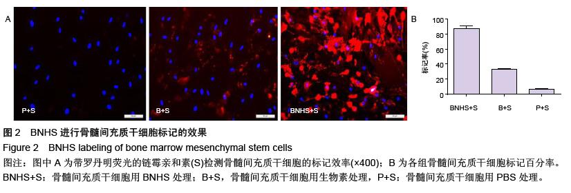
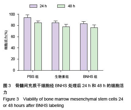
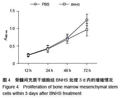
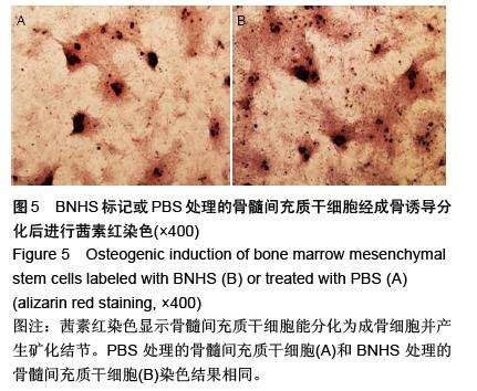
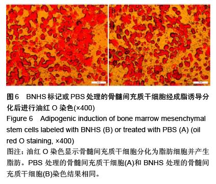
方法:全骨髓培养法得到第3代骨髓间充质干细胞,采用流式细胞仪鉴定;以磺化生物素-N-羟基琥珀酰亚胺、生物素、链霉亲和素将黏附分子配体唾液酸化的路易斯抗原装备到骨髓间充质干细胞表面;通过荧光显微镜评估骨髓间充质干细胞表面标记的效率,锥虫蓝染色法检测骨髓间充质干细胞的活性,CCK-8比色法检测骨髓间充质干细胞的增殖功能;成脂、成骨诱导检测骨髓间充质干细胞多分化功能。

结果与结论:①全骨髓培养法培养2周,可得到第3代骨髓间充质干细胞,细胞表达CD90,CD29,不表达CD34和CD45。②以生物素及链霉亲和素成功将黏附分子配体唾液酸LewisX(SleX)装备到骨髓间充质干细胞表面,且对细胞活性、增殖、分化功能影响不大。③运用这种方法对细胞进行表型修饰,操作技术简单,修改效率可达88%,有望提高骨髓间充质干细胞的归巢率,未来会有广泛和重要的应用价值。 

ORCID: 0000-0002-9556-3302 (杨林)

关键词: 干细胞, 骨髓干细胞, 生物素-链霉亲和素, 骨髓间充质干细胞, 磺化生物素- N-羟基琥珀酰亚胺, 唾液酸化的路易斯抗原, 国家自然科学基金

Abstract:

BACKGROUND: Currently, there is a lack of efficient, non-invasive way to transplant stem cells to the target organ or tissue. Exploring a way to guide targeting transplantation of stem cells and to improve the efficiency of stem cell homing is now one of focuses in the field of stem cells research.
OBJECTIVE: To establish a simple and feasible method to chemically modify the cell surface using biotin-streptavidin reaction system, and to evaluate the efficiency of this method to label bone marrow mesenchymal stem cells (BMSCs) and its effects on cell biological functions.
METHODS: Passage 3 BMSCs were obtained by whole bone marrow culture method and verified by flow cytometry. Biotin, streptavidin, sulfonated biotin-N-hydroxy-succinimide were used to equip the adhesion molecule ligand, sialyated LewisX (SLeX), to the BMSCs surface. The labeling rate of BMSCs was assessed using fluorescence microscope, the vitality of BMSCs was evaluated by trypan blue staining, and the proliferation of BMSCs was evaluated by cell counting kit-8 assay. Adipogenic and osteogenic inductions were used to evaluate the effect of the method on the multi-differentiation function of BMSCs.
RESULTS AND CONCLUSION: After culture for 2 weeks, passage 3 BMSCs were obtained and confirmed by expressing CD90, CD29 and lack of CD34, CD45. Biotin, streptavidin, sulfonated biotin-N-hydroxy-succinimide were successfully used to equip sialyated LewisX (SLeX) to the BMSCs surface and had minor effects on the vitality, proliferation, and differentiation of BMSCs. This method was simple for surface modification and had a high modification rate of 88%. The homing of BMSCs modified by this method to the target organ or tissue could be greatly enhanced. Therefore, this method potentially could have extensive and important applications.