| [1] 李丽.1000例儿童口腔内科疾病的回顾性分析[J].中国医学创新,2009,6(18):37.
[2] 樊明文.牙体牙髓病学[M].2版.北京:人民卫生出版社, 2003: 151-152.
[3] Cao H,Qi Z,Jiang H,et al.Detection of Porphyromonas endodontalis,Porphyromonas gingivalis and Prevotella intermedia in primary endodontic infections in a Chinese population.Int Endod J.2012;45(8):773-781.
[4] 李任,仇丽鸿.牙髓卟啉单胞菌内毒素致病性研究进展[J].中国实用口腔医学杂志,2009,2(12):756-758.
[5] 张震康,俞光岩.实用口腔医学[M].3版.北京:人民卫生出版社, 2009:59.
[6] Dumani A,Yoldas O,Yilmas S,et al.Polymerase chain reaction of enterococcus faecalis and candida albicans in apical periodontitis from Turkish patients.J Clin Exp Dent. 2012; 4(1):e34-e39.
[7] Murad CF,Sassone LM,Faveri M,et al.Microbial diversity in persistent root canal infections investigated by checkerboard DNA-DNA hybridization.J Endod.2014;40(7):899-906.
[8] Yang QB,Fan LN,Shi Q.Polymerase Chain Reaction-Denaturing Gradient Gel Electrophoresis, Cloning,and Sequence Analysis of Bacteria Associated with Acute Periapical Abscesses in Children.J Endod. 2010;36(2): 221-223.
[9] Shang JJ,Yang QB,Zhao HY,et al.Preliminary molecular analysis of bacterial composition in periapical lesions with primary endodontic infections of deciduous teeth.Chin Med J.2013;126(16):3112-3117.
[10] Hess D,Solomon E,Spears R,et al. Retreatability of a bioceramic root canal sealing material.J Endod. 2011; 37(11):1547-1549.
[11] Loushine BA,Bryan TE,Looney SW,et al.Setting properties and cytotoxicity evaluation of a premixed bioceramic root canal sealer. J Endod.2011;37(5):673-677.
[12] Damas BA,Wheater MA,Bringas JS,et al. Cytotoxicity comparison of mineral trioxide aggregates and EndoSequence bioceramic root repair materials.J Endod. 2011;37(3):372-375.
[13] Leal F,De-Deus G,Brandao C,et al. Comparison of the root-end seal provided by bioceramic repair cements and White MTA.Int Endod.2011;44(7):662-668.
[14] Chandra SS,Shankar P,Indira R.Depth of penetration of four resin sealers into radicular dentinal tubules:a confocal microscopic study.J Endod.2012;38(10):1412-1416.
[15] Candeiro GT,Correia FC,Duarte MA,et al.Evaluation of radiopacity,pH,release of calcium ions,and flow of a bioceramic root canal sealer.J Endod.2012;38(6):842-845.
[16] Ersahan S,Aydin C.Solubility and apical sealing characteristics of a new calcium silicate-based root canal sealer in comparison to calcium hydroxide-,methacrylate resin- and epoxy resin-based sealers.Acta Odontol Scand. 2013;71(3-4):857-862.
[17] 王芬,薛明.镍钛器械的研究进展[J].中国实用口腔科杂志, 2011,4(6):335-340.
[18] Keles A,Ahmetoglu F,Simsek N,et al.Heat conductive properties of set root canal sealers.Acta Odontol Scand. 2013;71(3-4):751-755.
[19] 房俊艳,贾云飞,宋永海,等. 根管封闭剂封闭性能检测方法研究进展[J].当代医学,2011,17(3):34-35.
[20] Ma J,Sben Y,Stojicic S,et al.Biocompatibility of two novel root repair materials.J Endod,2011;37(6):793-798.
[21] 徐悦,赵云富,周洁.根管充填材料根尖微渗漏的评估方法研究进展[J].口腔医学,2010,30(8):502-504.
[22] Mollo A,Botti G,Prinicipi Goldoni N,et al.Efficacy of two Ni-Ti systems and hand files for removing gutta-percha from root canals.Int Endod J.2012;45(1):1-6.
[23] Betti LV,Bramante CM,de Moraes LG,et al.Comparison of GPX with or without solvent and hand files in removing filling Materials from root canals-An exvivo study.Oral Surg Oral Med Oral Pathol.2010;110(5):685 -680.
[24] 郭晓琳,彭伟,鲁丽珍,等.不同根管预备器械对根管充填后冠方微渗漏影响的实验研究[J].牙体牙髓牙周病学杂志, 2012, 22 (10): 579-581.
[25] 杨涛.不同根管预备方法和不同根管充填技术组合对根尖微渗漏的影响[J].牙体牙髓牙周病学杂志,2013,23(7):458-461.
[26] 刘智永,盛芳,郝新宇,等.不同根管预备技术对弯曲根管封闭性能的影响[J].山东大学学报(医学版),2010,48(9):68-71.
[27] 范兵,樊明文,边专,等.根管偏移对充填材料封闭根管能力的影响[J].口腔医学纵横,2001,17(2):83-87.
[28] 张琛,黄巍,侯本祥.4种根管充填技术的根尖微渗漏评价[J].北京口腔医学, 2010,18(6):319-322.
[29] ]赵军.Thermafil充填技术和冷侧压法的根管微渗漏研究[J].当代医学,2008,12(12):142.
[30] 莫清波,陈桂军.Thermafil根管充填技术的临床研究[J].华夏学, 2010,23(3):306-307.
[31] 吴俊,彭彬,范兵,等.Thermafil充填技术体外实验研究[J].牙体牙髓牙周病学杂志,2003,13(3):149-151.
[32] 赵岩,慕红文.三种根管充填方法根尖微渗漏的实验研究[J].天津医科大学学报,2010,16(4):659-661.
[33] 王冬梅,沈嵩,高学军.根管细菌渗漏体外模型的建立[J].牙体牙髓牙周病学杂志,2006,16(5):250-253.
[34] 徐琼,樊明文,范兵.葡萄糖定量分析根管微渗漏模型的建立[J].现代口腔医学杂志,2003,17(3):215-217.
[35] 贡艳宏,张光东,刘卫红,等.采用葡萄糖定量分析法评价连续波牙胶充填技术的根管封闭性能的研究[J].口腔医学, 2012,32 (11): 646-648.
[36] 吴雪,宫佳乐,王效军,等.MTA及GIC对MG-63细胞增殖和分化功能影响的实验研究[J].口腔医学研究,2011,27(11):960-962.
[37] 刘阗,周会喜,徐平平,等.MTA和Vitapex糊剂治疗成人慢性根尖周炎伴根尖孔闭合不全的疗效比较[J].口腔医学研究, 2010, 26 (3):400-402.
[38] 张林祺,刘双云,张昀,等.MTA用于根端囊肿根尖倒充填的临床评价[J].口腔颌面外科杂志,2013,23(5):382-383.
[39] Song M,Kim E.A prospective randomized controlled study of mineral trioxide aggregate and super ethoxybenzoic acid as root-end filling materials in endodontic microsurgery.J Endod. 2012;38(7):875-879.
[40] 单海琴.三氧化矿物凝聚体修补髓室底穿孔的疗效观察[J].实用口腔医学杂志,2010,26(3):420-421.
[41] 张莹,唐荣银,麻明歌,等.用MTA修复髓室底较大穿孔的临床观察[J].牙体牙髓牙周病学杂志,2008,18(7):386-389.
[42] 陈超,肖海波.MTA与GuttaFlow封闭根管效果的对比研究[J].医学临床研究,2010,27(11):2128-2129.
[43] Gandolfi MQ,Prati C.MTA and F-doped MTA cements used as sealers with warm gutta-percha.Long-term study of sealing ability.Int Endod J.2010;31(7):57-59.
[44] AsgaryS,Eghbal MJ,Ehsani S.Periradicular regeneration after endodontic surgery with calcium-enriched mixture cemerit in dogs.J Endod.2010;36(5):837-841. |
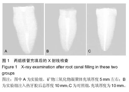
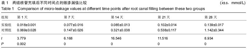
.jpg)
.jpg)