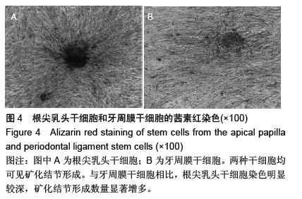
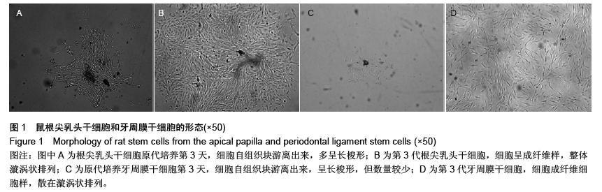
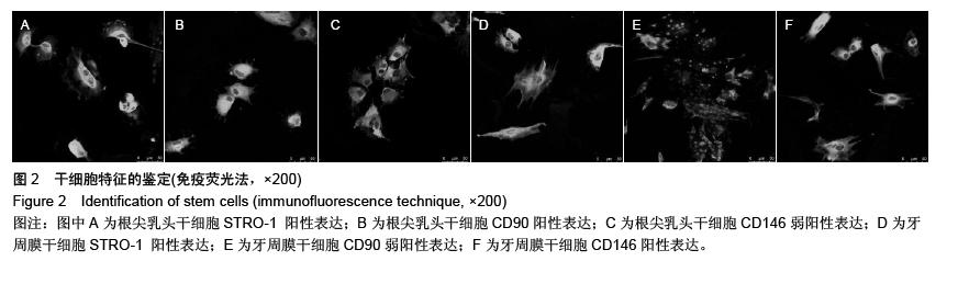
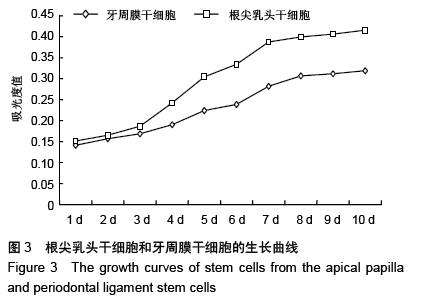
| [1] Sonoyama W, Liu Y, Fang D, et al. Mesenchymal stem cell-mediated functional tooth regeneration in swine. PLoS One. 2006;1:e79.[2] Sonoyama W, Liu Y, Yamaza T, et al. Characterization of the apical papilla and its residing stem cells from human immature permanent teeth: a pilot study. J Endod. 2008; 34(2):166-171.[3] Abe S, Yamaguchi S, Watanabe A, et al. Hard tissue regeneration capacity of apical pulp derived cells (APDCs) from human tooth with immature apex. Biochem Biophys Res Commun. 2008;371(1):90-93.[4] Bakopoulou A, Leyhausen G, Volk J, et al. Comparative analysis of in vitro osteo/odontogenic differentiation potential of human dental pulp stem cells (DPSCs) and stem cells from the apical papilla (SCAP). Arch Oral Biol. 2011;56(7):709-721.[5] Huang GT, Sonoyama W, Liu Y, et al. The hidden treasure in apical papilla: the potential role in pulp/dentin regeneration and bioroot engineering.J Endod. 2008;34(6):645-651.[6] 熊华翠,陈柯,黄义彬,等.人根尖乳头干细胞生成牙髓牙本质复合体的实验研究[J]. 南方医科大学学报,2013,33(10):1512-1516.[7] 中华人民共和国科学技术部.关于善待实验动物的指导性意见. 2006-09-30.[8] 司徒镇强,吴军正.细胞培养[M].北京:世界图书出版社,2007.[9] 王燕萍,吴锦涛,王子露,等.磷酸二氢钾对根尖牙乳头干细胞成牙及成骨向分化能力的影响[J].中华口腔医学杂志, 2013, 48(1): 27-31.[10] 高润涛,范志朋.过表达BCOR基因抑制根尖牙乳头干细胞成肌分化[J].口腔生物医学, 2013, 4(1):1-3.[11] 杜鹃,范志朋.NFkB信号通路在炎症根尖牙乳头干细胞中的作用[J].北京口腔医学, 2013, 21(1):9-12.[12] 刁树,杨东梅,范志朋.牙组织源性干细胞的微环境[J]. 中华口腔医学杂志, 2014, 49(4):254-256.[13] 姚睿,范志朋.组蛋白去甲基化酶KDM4B促进根尖牙乳头干细胞中成骨和成牙本质分化[J]. 北京口腔医学, 2013, 21(4): 181-184.[14] 杨海兵,韩萱,杨琳.血管内皮生长因子和转化因子β1基因调控人根尖乳头细胞矿化相关因子的研究[J]. 华西口腔医学杂志, 2012, 30(5):468-472. [15] 邱云,郑青,萧树东,等.干细胞微环境的体外模拟[J]. 中国组织工程研究与临床康复, 2011, 15(6):1123-1126.[16] 吴家媛,贾谦,李帅,等.MTA对人根尖牙乳头干细胞体外增殖的影响[J]. 齐齐哈尔医学院学报,2011,32(15):2400-2401.[17] Banchs F, Trope M. Revascularization of immature permanent teeth with apical periodontitis: new treatment protocol. J Endod. 2004;30(4):196-200.[18] Chueh LH, Huang GT. Immature teeth with periradicular periodontitis or abscess undergoing apexogenesis: a paradigm shift. J Endod. 2006;32(12):1205-1213.[19] Cotti E, Mereu M, Lusso D. Regenerative treatment of an immature, traumatized tooth with apical periodontitis: report of a case. J Endod. 2008;34(5):611-616.[20] Ding RY, Cheung GS, Chen J, et al. Pulp revascularization of immature teeth with apical periodontitis: a clinical study. J Endod. 2009;35(5):745-749.[21] Petrino JA, Boda KK, Shambarger S, et al. Challenges in regenerative endodontics: a case series. J Endod. 2010;36(3): 536-541.[22] Chen MY, Chen KL, Chen CA, et al. Responses of immature permanent teeth with infected necrotic pulp tissue and apical periodontitis/abscess to revascularization procedures. Int Endod J. 2012;45(3):294-305.[23] Jung IY, Kim ES, Lee CY, et al. Continued development of the root separated from the main root. J Endod. 2011;37(5): 711-714.[24] Huang GT, Yamaza T, Shea LD, et al. Stem/progenitor cell-mediated de novo regeneration of dental pulp with newly deposited continuous layer of dentin in an in vivo model. Tissue Eng Part A. 2010;16(2):605-615.[25] Gronthos S, Mankani M, Brahim J, et al. Postnatal human dental pulp stem cells (DPSCs) in vitro and in vivo. Proc Natl Acad Sci U S A. 2000;97(25):13625-13630.[26] 武曦,张纲,谭颖徽. Notch信号通路在牙髓干细胞增殖和分化中的调控作用[J]. 牙体牙髓牙周病学杂志, 2011, 21(5):298-302.[27] Morsczeck C, Götz W, Schierholz J, et al. Isolation of precursor cells (PCs) from human dental follicle of wisdom teeth. Matrix Biol. 2005;24(2):155-165.[28] 高东辉,李军,孙晶,等.牙周膜干细胞的研究进展[J].中国老年学杂志,2012,32(15): 3362-3364.[29] Seo BM, Miura M, Gronthos S, et al. Investigation of multipotent postnatal stem cells from human periodontal ligament. Lancet. 2004;364(9429):149-155.[30] 熊华翠.人根尖乳头干细胞与牙周膜干细胞体外生物学特性的比较研究[D].广州:南方医科大学,2013. [31] 鲁少文, 税艳青. 牙周膜干细胞的研究进展[J]. 国际口腔医学杂志, 2013, 40(6): 769-772.[32] 孙静.牙周膜干细胞巢与牙周组织再生[J]. 国际口腔医学杂志, 2011, 38(4): 460-462.[33] 马丽, 杨丕山, 王燕. 人牙根尖乳头细胞的培养及TNF-α 对其增殖的影响[J].上海口腔医学, 2010, 19(5): 525-529.[34] 刘彩奇,陈柯,黄义彬,等. TNF-α 对人根尖乳头干细胞增殖及分化能力的影响[J].口腔医学研究, 2014, 30(5): 392-395. [35] Huang GT. Pulp and dentin tissue engineering and regeneration: current progress. Regen Med. 2009;4(5): 697-707.[36] 黄义彬,陈柯,熊华翠.牙根持续发育期根尖乳头干细胞的研究进展[J].口腔医学研究, 2013, 29(5): 490-492.[37] 郭俊,杨健.人根尖牙乳头细胞分离、培养的研究[J].口腔医学研究, 2011, 27(11): 1010-1012. [38] 张瑛,宋莉.牙周膜干细胞的研究进展[J].中国组织工程研究与临床康复, 2011, 15(36): 6817-6820.[39] 董正谋,刘鲁川.牙周膜干细胞的研究进展[J].国际口腔医学杂志, 2012, 39(4): 519-522.[40] 郭俊,杨健.人根尖乳头干细胞及其在组织工程中的研究进展[J]. 国际口腔医学杂志, 2010, 37(5): 464-466. |
.jpg)
.jpg)