| [1] 陈业刚,刘晓强.肾癌患者循环肿瘤细胞的研究进展[J].中国肿瘤临床,2011,38(3):175-178.
[2] 胡峻珲,叶章群.肾脏肿瘤的实验研究现状和进展[J].中华实验外科杂志,2013,30(6):1107-1109.
[3] 周天贵,周承贵,陈先国,等.肾癌组织中MMP-7表达和MVD测定及其临床意义[J].山东医药,2008,48(40):22-24.
[4] 张朝华,黄研,彭红涛,等.肾癌免疫治疗研究新进展[J].中国药业,2012,21(2):79-80.
[5] 蒋焜,郑新民. 肾癌的治疗进展--分子靶向治疗[J].医学新知杂志,2014,24(2):79-81.
[6] 周世阳, 陈伟. MSCT对肾癌诊断与分期的临床意义[J].重庆医学,2012,41(19):1989-1991.
[7] Singh SK, Clarke ID, Hide T, et al. Cancer stem cells in nervous system tumors. Oncogene. 2004;23(43):7267-7273.
[8] Yu Z, Pestell TG, Lisanti MP, et al. Cancer stem cells. Int J Biochem Cell Biol. 2012;44(12):2144-2151.
[9] Visvader JE, Lindeman GJ. Cancer stem cells: current status and evolving complexities.Cell Stem Cell. 2012;10(6):717-728.
[10] Bonnet D, Dick JE. Human acute myeloid leukemia is organized as a hierarchy that originates from a primitive hematopoietic cell. Nat Med. 1997;3(7):730-737.
[11] Ponti D, Costa A, Zaffaroni N, et al. Isolation and in vitro propagation of tumorigenic breast cancer cells with stem/progenitor cell properties. Cancer Res. 2005;65(13): 5506-5511.
[12] Zhang S, Balch C, Chan MW, et al. Identification and characterization of ovarian cancer-initiating cells from primary human tumors. Cancer Res. 2008;68(11):4311-4320.
[13] Horta S, Agostinho AL, Mateus R, et al. Looking out for cancer stem cells' properties: the value-driving role of CD44 for personalized medicines. Curr Cancer Drug Targets. 2015; 14(9):832-849.
[14] Li C, Heidt DG, Dalerba P, et al. Identification of pancreatic cancer stem cells. Cancer Res. 2007;67(3):1030-1037.
[15] Qin J, Liu X, Laffin B, et al. The PSA(-/lo) prostate cancer cell population harbors self-renewing long-term tumor- propagating cells that resist castration. Cell Stem Cell. 2012;10(5):556-569.
[16] Bussolati B, Bruno S, Grange C, et al. Identification of a tumor-initiating stem cell population in human renal carcinomas. FASEB J. 2008;22(10):3696-3705.
[17] Bussolati B, Dekel B, Azzarone B, et al. Human renal cancer stem cells. Cancer Lett. 2013;338(1):141-146.
[18] Wang L, Park P, La Marca F, et al. BMP-2 inhibits tumor-initiating ability in human renal cancer stem cells and induces bone formation. J Cancer Res Clin Oncol. 2015; 141(6):1013-1024.
[19] Nishizawa S, Hirohashi Y, Torigoe T, et al. HSP DNAJB8 controls tumor-initiating ability in renal cancer stem-like cells. Cancer Res. 2012;72(11):2844-2854.
[20] Gassenmaier M, Chen D, Buchner A, et al. CXC chemokine receptor 4 is essential for maintenance of renal cell carcinoma-initiating cells and predicts metastasis. Stem Cells. 2013;31(8):1467-1476.
[21] Zhu X, Fang L, Du G, et al. Effects of Danshen on the Cardiovascular System. Dan Shen (Salvia miltiorrhiza) in Medicine: Springer, 2015:79-127.
[22] Zhou L, Zuo Z, Chow MS. Danshen: an overview of its chemistry, pharmacology, pharmacokinetics, and clinical use. J Clin Pharmacol. 2005;45(12):1345-1359.
[23] Wojcikowski K, Johnson DW, Gobe G. Herbs or natural substances as complementary therapies for chronic kidney disease: ideas for future studies. J Lab Clin Med. 2006; 147(4):160-166.
[24] Wang BE. Treatment of chronic liver diseases with traditional Chinese medicine. J Gastroenterol Hepatol. 2000;15 Suppl: E67-70.
[25] Yoo KY, Park SY. Terpenoids as potential anti-Alzheimer's disease therapeutics. Molecules. 2012;17(3):3524-3538.
[26] Li W, Saud SM, Young MR, et al. Cryptotanshinone, a Stat3 inhibitor, suppresses colorectal cancer proliferation and growth in vitro. Mol Cell Biochem. 2015;406(1-2):63-73.
[27] Xu D, Lin TH, Li S, et al. Cryptotanshinone suppresses androgen receptor-mediated growth in androgen dependent and castration resistant prostate cancer cells. Cancer Lett. 2012;316(1):11-22.
[28] Li S, Wang H, Hong L, et al. Cryptotanshinone inhibits breast cancer cell growth by suppressing estrogen receptor signaling. Cancer Biol Ther. 2015;16(1):176-184.
[29] Zhang YF, Zhang M, Huang XL, et al. The combination of arsenic and cryptotanshinone induces apoptosis through induction of endoplasmic reticulum stress-reactive oxygen species in breast cancer cells. Metallomics. 2015;7(1):165-173.
[30] Chen W, Lu Y, Chen G, et al. Molecular evidence of cryptotanshinone for treatment and prevention of human cancer. Anticancer Agents Med Chem. 2013;13(7):979-987.
[31] Chen L, Wang HJ, Xie W, et al. Cryptotanshinone inhibits lung tumorigenesis and induces apoptosis in cancer cells in vitro and in vivo. Mol Med Rep. 2014;9(6):2447-2452.
[32] Park IJ, Yang WK, Nam SH, et al. Cryptotanshinone induces G1 cell cycle arrest and autophagic cell death by activating the AMP-activated protein kinase signal pathway in HepG2 hepatoma. Apoptosis. 2014;19(4):615-628.
[33] Soeda A, Park M, Lee D, et al. Hypoxia promotes expansion of the CD133-positive glioma stem cells through activation of HIF-1alpha. Oncogene. 2009;28(45):3949-3959.
[34] Singh SK, Hawkins C, Clarke ID, et al. Identification of human brain tumour initiating cells. Nature. 2004;432(7015):396-401.
[35] 吴恭瑾,卢建忠,杨宁强,等.免疫磁珠法分离纯化人肾癌CD133+细胞实验研究[J].甘肃医药,2012,31(8):561-564.
[36] 樊胜玉.肾癌的诊断和手术治疗研究进展[J].中国民康医学, 2013, 25(10):108-109.
[37] 李伟,李晓宇,黄建华,等.生物信息学方法分析人肾细胞癌中潜在的miRNAs生物标志物和其调控的关键通路[J].临床泌尿外科杂志,2015,30(6):535-540.
[38] Escudier B, Porta C, Schmidinger M, et al. Renal cell carcinoma: ESMO Clinical Practice Guidelines for diagnosis, treatment and follow-up. Ann Oncol. 2014;25 Suppl 3:iii49-56.
[39] 弓建华,陈春雷.隐丹参酮抗肿瘤作用的研究进展[J].广东医学院学报,2012,30(1):87-90.
[40] 叶欢,阮君山,王少明.隐丹参酮抗肿瘤转移的研究进展[J].中国药理学通报,2014,30(7):893-896.
[41] Chen W, Luo Y, Liu L, et al. Cryptotanshinone inhibits cancer cell proliferation by suppressing Mammalian target of rapamycin- mediated cyclin D1 expression and Rb phosphorylation. Cancer Prev Res (Phila). 2010;3(8):1015-1025.
[42] Shin DS, Kim HN, Shin KD, et al. Cryptotanshinone inhibits constitutive signal transducer and activator of transcription 3 function through blocking the dimerization in DU145 prostate cancer cells. Cancer Res. 2009;69(1):193-202.
[43] Chen W, Liu L, Luo Y, et al. Cryptotanshinone activates p38/JNK and inhibits Erk1/2 leading to caspase-independent cell death in tumor cells. Cancer Prev Res (Phila). 2012;5(5):778-787.
[44] Jung JH, Kwon TR, Jeong SJ,et al. Apoptosis Induced by Tanshinone IIA and Cryptotanshinone Is Mediated by Distinct JAK/STAT3/5 and SHP1/2 Signaling in Chronic Myeloid Leukemia K562 Cells. Evid Based Complement Alternat Med. 2013;2013:805639.
[45] 叶因涛,徐文清,仲巍.隐丹参酮对宫颈癌Hela细胞增殖及细胞凋亡的影响[J].中国中药杂志,2010,35(1):118-121. |
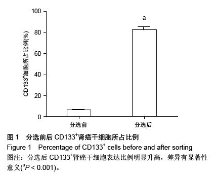
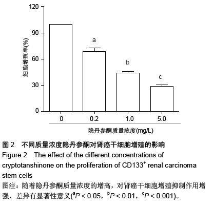
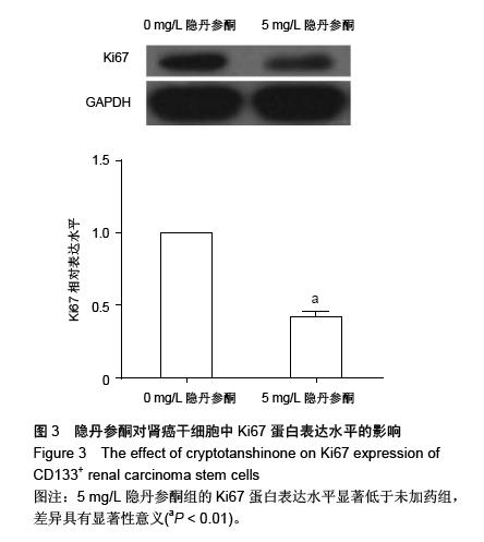
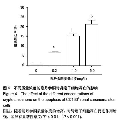
.jpg)