| [1] Richards M, Huibregtse BA, Caplan AI, et al. Marrow-derived progenitor cell injections enhance new bone formation during distraction. J Orthop Res. 1999;17(6):900-908.
[2] Ouyang JF, Lou J, Yan C, et al. In-vitro promoted differentiation of mesenchymal stem cells towards hepatocytes induced by salidroside. J Pharm Pharmacol. 2010;62(4):530-538.
[3] Lu P, Blesch A, Tuszynski MH. Induction of bone marrow stromal cells to neurons: differentiation, transdifferentiation, or artifact. J Neurosci Res. 2004;77(2):174-191.
[4] Cipriani P, Guiducci S, Miniati I, et al. Impairment of endothelial cell differentiation from bone marrow-derived mesenchymal stem cells: new insight into the pathogenesis of systemic sclerosis. Arthritis Rheum. 2007;56(6):1994-2004.
[5] Clabaut A, Delplace S, Chauveau C, et al. Human osteoblasts derived from mesenchymal stem cells express adipogenic markers upon coculture with bone marrow adipocytes. Differentiation. 2010;80(1):40-45.
[6] Fukuda K. Molecular characterization of regenerated cardiomyocytes derived from adult mesenchymal stem cells. Congenit Anom (Kyoto). 2002;42(1):1-9.
[7] Makino S, Fukuda K, Miyoshi S, et al. Cardiomyocytes can be generated from marrow stromal cells in vitro. J Clin Invest. 1999;103(5):697-705.
[8] Woodbury D, Reynolds K, Black IB. Adult bone marrow stromal stem cells express germline, ectodermal, endodermal, and mesodermal genes prior to neurogenesis. J Neurosci Res. 2002;69(6):908-917.
[9] Oh SH, Muzzonigro TM, Bae SH, et al. Adult bone marrow-derived cells trans-differentiating into insulin-producing cells for the treatment of type I diabetes. Lab Invest. 2004;84(5):607-617.
[10] 李秀华,张雷,王海萍,等. P19 细细胞体外DMSO诱导向心肌样细胞分化[J].第四军医大学学报, 2006, 27(2):129-132.
[11] Sotkis HV, Lazarenko PM, Boldyriev OI, et al. Identification of E-4031-sensitive potassium current component in murine P19 embryonic carcinoma cell line differentiated in cardiomyocytes. Fiziol Zh. 2006;52(1):49-61.
[12] Moscoso I, Centeno A, López E, et al. Differentiation "in vitro" of primary and immortalized porcine mesenchymal stem cells into cardiomyocytes for cell transplantation. Transplant Proc. 2005;37(1):481-482.
[13] Fukuda K. Development of regenerative cardiomyocytes from mesenchymal stem cells for cardiovascular tissue engineering. Artif Organs. 2001;25(3):187-193.
[14] Krysko DV, Vanden Berghe T, D'Herde K, et al. Apoptosis and necrosis: detection, discrimination and phagocytosis. Methods. 2008;44(3):205-221.
|
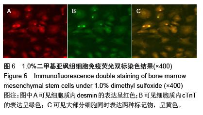
.jpg)
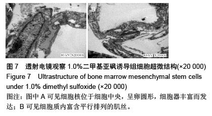
.jpg)
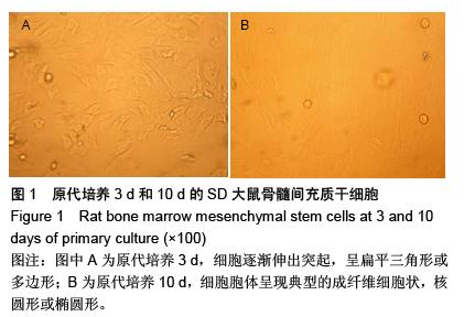
.jpg)
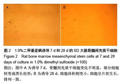
.jpg)