| [1] Martin I, Miot S,Barbero A,et al.Osteochondral tissue engineering. J Biomech.2007; 40:750-765.
[2] Nukavarapu SP,Dorcemus DL.Osteochondral tissue engineering: Current strategies and challenges.Biotechnol Adv.2013;31:706-721.
[3] Horner EA,Kirkham J,Wood D,et al.Long bone defect models for tissue engineering applications: criteria for choice.Tissue Eng Part B Rev.2010;16:263-271.
[4] Melton JT,Wilson AJ,Chapman-Sheath P,et al.bone plug: chondral repair, scaffold design, surgical technique and early experiences.Expert Rev Med Devices.2010;7:333-341.
[5] Omenetto FG,Kaplan DL.New opportunities for an ancient material.Science.2010;329: 528-531.
[6] Kundu B,Rajkhowa R,Kundu SC,et al.Silk fibroin biomaterials for tissue regenerations.Adv Drug Deliv Rev.2013;65: 457-470.
[7] Velema J,Kaplan D.Biopolymer-based biomaterials as scaffolds for tissue engineering.Adv Biochem Eng Biotechnol. 2006;102:187-238.
[8] Wray LS,Rnjak-Kovacina J,Mandal BB,et al.A silk-based scaffold platform with tunable architecture for engineering critically-sized tissue constructs. Biomaterials. 2012;33: 9214-9224.
[9] Rodrigues MT,Gomes ME,Reis RL.Current strategies for osteochondral regeneration: from stem cells to pre-clinical approaches.Curr Opin Biotechnol.2011;22(5):726-733.
[10] Patra C,Talukdar S,Novoyatleva T,et al.Silk protein fibroin from Antheraea mylitta for cardiac tissue engineering. Biomaterials.2012;33:2673-2680.
[11] Rockwood DN,Preda RC,Yucel T,et al.Materials fabrication from Bombyx mori silk fibroin.Nat Protoc.2011;6:1612-1631.
[12] Saha S,Kirkham J,Wood D,et al.Comparative study of the chondrogenic potential of human bone marrow stromal cells, neonatal chondrocytes and adult chondrocytes.Biochem Biophys Res Commun.2010;401:333-338.
[13] Mandal BB,Grinberg A,Seok Gil E,et al.High-strength silk protein scaffolds for bone repair.PNAS.2012;109:7699-7704.
[14] Vunjak-Novakovic G,Obradovic B,Martin I,et al.Dynamic cell seeding of polymer scaffolds for cartilage tissue engineering. Biotechnol Prog.1998;14:193-202.
[15] Bhardwaj N,Kundu SC.Chondrogenic differentiation of rat MSCs on porous scaffolds of silk fibroin/chitosan blends. Biomaterials.2012;33:2848-2857.
[16] Shao X,Goh JC,Hutmacher DW,et al.Repair of large articular osteochondral defects using hybrid scaffolds and bone marrow-derived mesenchymal stem cells in a rabbit model. Tissue Eng.2006;12(6):1539-1551.
[17] Seda T??l? R,Ghosh S,Laha MM,et al.Comparative chondrogenesis of human cell sources in 3D scaffolds.J Tissue Eng Regen Med.2009;3(5):348-360.
[18] Hunziker EB.Articular cartilage repair: basic science and clinical progress. A review of the current status and prospects.Osteoarthritis Cartilage.2002;10(6):432-463.
[19] Shanmugasundaram S,Chaudhry H,Arinzeh TL.Microscale versus nanoscale scaffold architecture for mesenchymal stem cell chondrogenesis.Tissue Eng Part A.2011;17(5-6):831-840.
[20] Stenhamre H,Nannmark U,Lindahl A,et al.Brittberg M Influence of pore size on the redifferentiation potential of human articular chondrocytes in poly(urethane urea) scaffolds. J Tissue Eng Regen Med.2011;5(7):578-588.
[21] Im GI,Ko JY,Lee JH.Chondrogenesis of adipose stem cells in a porous polymer scaffold: influence of the pore size.Cell Transplant.2012;21(11):2397-2405.
[22] Mandal BB,Das S,Choudhury K,et al.Implication of silk film RGD availability and surface roughness on cytoskeletal organization and proliferation of primary rat bone marrow cells.Tissue Eng Part A.2010;16(7):2391-2403.
[23] Yang HS,La WG,Bhang SH,et al.Apatite-Coated Collagen Scaffold for Bone Morphogenetic Protein-2 Delivery.Tissue Eng Part A.2011;17:2153-2156.
[24] Chen S,Xuetao S,Morita H,et al.BMP-2-loaded silica nanotube fibrous meshes for bone generation.Sci Technol Adv Mater.2011;12:065003.
[25] Meinel L,Betz O,Fajardo R,et al.Silk based biomaterials to heal critical sized femur defects.Bone.2006;39:922-931.
[26] Riccio M,Maraldi T,Pisciotta A,et al.Fibroin scaffold repairs critical-size bone defects in vivo supported by human amniotic fluid and dental pulp stem cells.Tissue Eng Part A.2012;18: 1006-1013. |
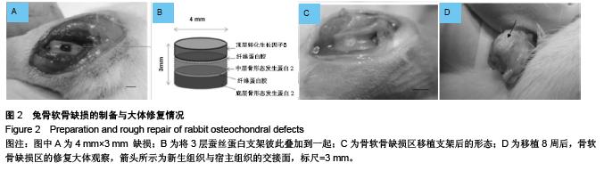
.jpg) 2.2 体内骨软骨修复区组织学观察结果 移植填充多层桑蚕丝蛋白支架及柞蚕丝蛋白的动物关节软骨缺损区情况见图2A-C。在支架移植后8周,移植各组缺损区均可见明显的界线(图2D)。大体观察,移植各组修复再生区表面都较光滑,覆盖着洁白的新生组织。显微镜下观察,实验1组和实验3组支架复合物内的细胞并没有至始至终维持其原有的形态(图3,4),而实验2组和实验4组的细胞始终维持其原来的形态(图5,6)。移植各组修复区域的细胞外基质都被天狼星红染料染成了特征性的红色,表明新生成的基质内胶原含量丰富。除实验1组外,阿新蓝染色阳性与葡萄糖胺聚糖集聚相一致。各实验组无论添加或不添加生长因子,顶层蚕丝蛋白支架上细胞的存在有利于封闭缺损表面,而空白对照组则缺损表面未能关闭。
2.2 体内骨软骨修复区组织学观察结果 移植填充多层桑蚕丝蛋白支架及柞蚕丝蛋白的动物关节软骨缺损区情况见图2A-C。在支架移植后8周,移植各组缺损区均可见明显的界线(图2D)。大体观察,移植各组修复再生区表面都较光滑,覆盖着洁白的新生组织。显微镜下观察,实验1组和实验3组支架复合物内的细胞并没有至始至终维持其原有的形态(图3,4),而实验2组和实验4组的细胞始终维持其原来的形态(图5,6)。移植各组修复区域的细胞外基质都被天狼星红染料染成了特征性的红色,表明新生成的基质内胶原含量丰富。除实验1组外,阿新蓝染色阳性与葡萄糖胺聚糖集聚相一致。各实验组无论添加或不添加生长因子,顶层蚕丝蛋白支架上细胞的存在有利于封闭缺损表面,而空白对照组则缺损表面未能关闭。
.jpg)
.jpg)
.jpg) 实验1组中胶原蛋白形成的量非常丰富,可见缺损区全层被染成红色(图3A)。实验2组可见支架内的胶原纤维排列整齐,在修复表面呈平行分布,在中间和底部区域呈垂直顶部的方向分布(图5A)。实验3组只在缺损修复区表面可见染成红色的胶原,支架复合物中间和底部主要是含有丰富葡萄糖胺聚糖的细胞外基质成分,被染成蓝色(图4A)。实验4组可见支架复合物表面和底部软骨样细胞有成团分布表现,而且只有此组细胞有这种分布情况(图6A)。通过阿新蓝/天狼星红染色图像合并方法对体内修复区进行分析,再次验证了组织学的发现(图3B,图4B,图5B,图6B)。所有移植实验组再生修复区底部都可见有血管组织生成(图3A,图4A,图5A,图6A)。空白对照组骨软骨缺损区在8周后都未能修复,表现为缺损区清晰可见,并且未能由细胞外基质完全填充。
实验1组中胶原蛋白形成的量非常丰富,可见缺损区全层被染成红色(图3A)。实验2组可见支架内的胶原纤维排列整齐,在修复表面呈平行分布,在中间和底部区域呈垂直顶部的方向分布(图5A)。实验3组只在缺损修复区表面可见染成红色的胶原,支架复合物中间和底部主要是含有丰富葡萄糖胺聚糖的细胞外基质成分,被染成蓝色(图4A)。实验4组可见支架复合物表面和底部软骨样细胞有成团分布表现,而且只有此组细胞有这种分布情况(图6A)。通过阿新蓝/天狼星红染色图像合并方法对体内修复区进行分析,再次验证了组织学的发现(图3B,图4B,图5B,图6B)。所有移植实验组再生修复区底部都可见有血管组织生成(图3A,图4A,图5A,图6A)。空白对照组骨软骨缺损区在8周后都未能修复,表现为缺损区清晰可见,并且未能由细胞外基质完全填充。.jpg)
.jpg)