中国组织工程研究 ›› 2015, Vol. 19 ›› Issue (43): 6940-6945.doi: 10.3969/j.issn.2095-4344.2015.43.010

• 细胞外基质材料 extracellular matrix materials • 上一篇    下一篇

脱细胞真皮基质修复猪胆管缺损:促进血管及胆管上皮再生

陈  刚,白建华,朱新锋,曹  俊,刘其雨,赵英鹏,李  立   

  1. 昆明市第一人民医院肝胆外科,云南省昆明市  650011
  • 收稿日期:2015-08-13 出版日期:2015-10-15 发布日期:2015-10-15
  • 通讯作者: 李立,博士,主任医师,昆明市第一人民医院肝胆外科,云南省昆明市 650011
  • 作者简介:陈刚,男,1969年生,四川省南充市人,汉族,博士,副主任医师,主要从事肝胆胰外科的基础与临床研究。

Acellular dermal matrix for repair of porcine bile duct defects: to promote vascular and bile duct epithelial regeneration

Chen Gang, Bai Jian-hua, Zhu Xin-feng, Cao Jun, Liu Qi-yu, Zhao Ying-peng, Li Li   

  1. Department of Hepatobiliary Surgery, the First People’s Hospital of Kunming, Kunming 650011, Yunnan Province, China
  • Received:2015-08-13 Online:2015-10-15 Published:2015-10-15
  • Contact: Li Li, M.D., Chief physician, Department of Hepatobiliary Surgery, the First People’s Hospital of Kunming, Kunming 650011, Yunnan Province, China
  • About author:Chen Gang, M.D., Associate chief physician, Department of Hepatobiliary Surgery, the First People’s Hospital of Kunming, Kunming 650011, Yunnan Province, China

摘要:

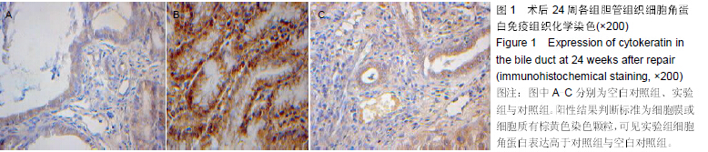
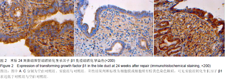
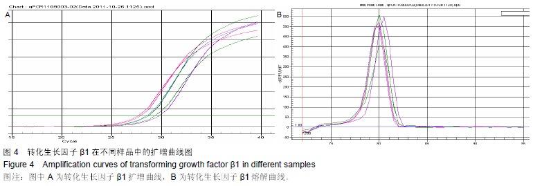
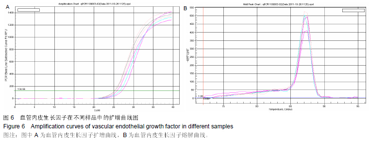
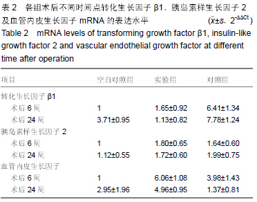
背景:脱细胞真皮基质是无细胞的天然组织支架,与人体软组织十分相近,易于塑形,无毒副作用,已被用于修补尿道与输尿管。
目的:观察脱细胞基质修补胆管损伤的效果。
方法:将30头滇南小耳猪随机均分为3组,空白对照组切断胆管后行端端吻合,实验组人为制作胆管缺损后以脱细胞真皮基质修补,对照组人为制作胆管缺损后以膨体聚四氟乙烯修补。修补后6,24周取胆管补片及其周围组织,进行免疫组织化学与RT-PCR检测。
结果与结论:免疫组织化学显示,实验组细胞角蛋白表达高于空白对照组与对照组,转化生长因子β1表达低于空白对照组与对照组。术后24周内RT-PCR检测显示,实验组总体转化生长因子β1基因表达低于空白对照组与对照组(P < 0.05),总体胰岛素样生长因子2基因表达高于空白对照组(P < 0.05),总体血管内皮生长因子基因表达高于空白对照组与对照组(P < 0.05)。表明脱细胞基质修复胆道损伤可促进血管及胆管上皮的再生,并且不增加瘢痕的形成。
中国组织工程研究杂志出版内容重点:生物材料;骨生物材料; 口腔生物材料; 纳米材料; 缓释材料; 材料相容性;组织工程

关键词: 生物材料, 材料相容性, 脱细胞基质, 胆管损伤, 胰岛素样生长因子2, 血管内皮生长因子A, 转化生长因子β1

Abstract:

BACKGROUND: Acellular dermal matrix is a cell-free natural tissue scaffold similar to human soft tissue, which is easy to shape and has non-toxic side effects. It has been used to repair the urethra and ureter.
OBJECTIVE: To investigate the effect of acellular dermal matrix on the repair of bile duct injury.
METHODS: Thirty Diannan miniature pigs were randomly divided into three groups: in blank group, the bile duct was resected followed by end to end anastomosis; in experimental group, bile duct defect model was made followed by repair with acellular dermal matrix; in control group, bile duct defect model was made followed by repair with expanded polytetrafluoroethylene. At 6 and 24 weeks after repair, bile duct patches and surrounding tissues were taken for immunohistochemical observation and RT-PCR detection.
RESULTS AND CONCLUSION: Compared with the control and blank group, the expression of cytokeratin was higher, but the expression of transforming growth factor β1 was lower in the experimental group. Within 24 weeks after repair, the total mRNA level of transforming growth factor β1 was lower in the experimental group than the other two groups (P < 0.05), but the total mRNA levels of insulin-like growth factor 2 and vascular endothelial growth factor were higher in the experimental group (P < 0.05). These findings indicate that the acellular dermal matrix for repair of bile duct injury can promote angiogenesis and bile duct epithelial regeneration, but not increase the formation of scars.
中国组织工程研究杂志出版内容重点:生物材料;骨生物材料; 口腔生物材料; 纳米材料; 缓释材料; 材料相容性;组织工程

Key words: Bile Ducts, Insulin-Like Growth Factor II, Transforming Growth Factor beta1, Tissue Engineering