中国组织工程研究 ›› 2015, Vol. 19 ›› Issue (41): 6659-6664.doi: 10.3969/j.issn.2095-4344.2015.41.018

• 干细胞移植 stem cell transplantation • 上一篇    下一篇

丙泊酚联合骨髓间充质干细胞移植对脊髓损伤大鼠后肢功能及电生理的影响

何 珏1,王天科2   

  1. 1赣南医学院病理学教研室,江西省赣州市 341000;2温州医科大学附属慈溪医院病理科,浙江省宁波市 315300
  • 出版日期:2015-10-01 发布日期:2015-10-01
  • 作者简介:女,1972年生,江西省赣州市人,汉族,1997年赣南医学院毕业,硕士,副教授,主要从事肿瘤病理学研究。

Propofol with bone marrow mesenchymal stem cell transplantation improves the hind limb function and electrophysiological changes in rats with spinal cord injury

He Jue1, Wang Tian-ke2   

  1. 1Department of Pathology, Gannan Medical University, Ganzhou 341000, Jiangxi Province, China; 2Department of Pathology, Affiliated Cixi Hospital of Wenzhou Medical University, Ningbo 315300, Zhejiang Province, China
  • Online:2015-10-01 Published:2015-10-01
  • About author:He Jue, Master, Associate professor, Department of Pathology, Gannan Medical University, Ganzhou 341000, Jiangxi Province, China

摘要:

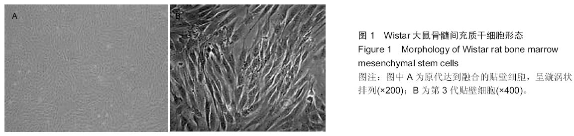
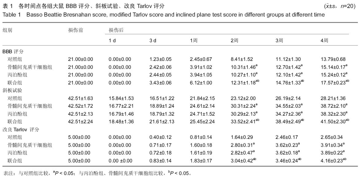
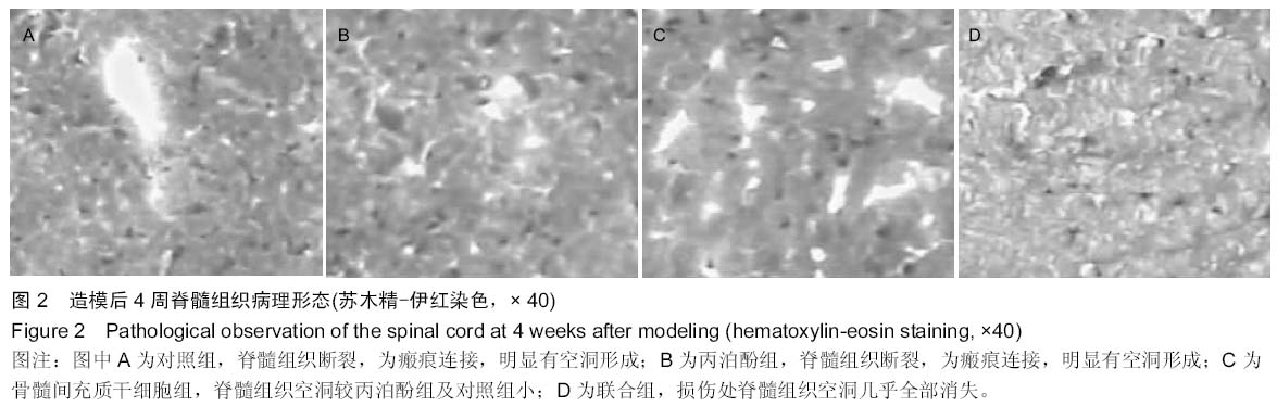
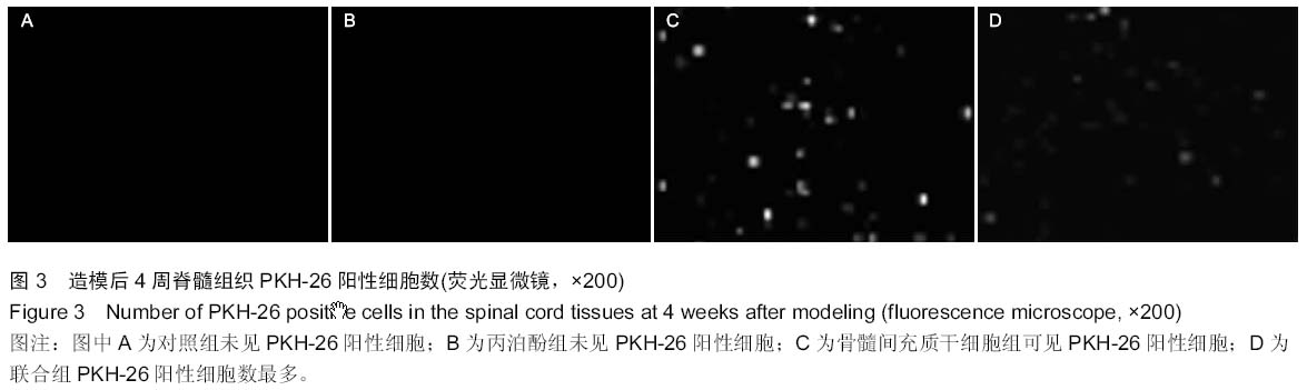
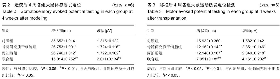
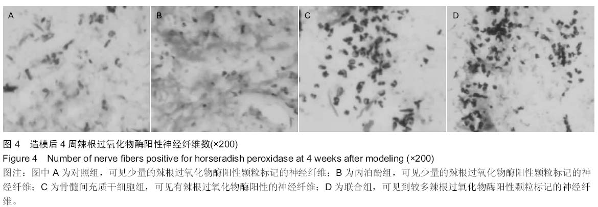
背景:研究发现,骨髓间充质干细胞具有修复神经元的作用,但单纯骨髓间充质干细胞移植对神经系统损伤的修复作用仍不理想。
目的:探讨骨髓间充质干细胞移植联合丙泊酚对脊髓损伤大鼠后肢功能和电生理的影响。
方法:80只成年的Wistar大鼠,建立脊髓损伤模型,按照随机数字表法分为4组(n=20):骨髓间充质干细胞组、对照组、联合组、丙泊酚组。建模后6 h通过1 mL注射器经尾静脉注入骨髓间充质干细胞悬液、细胞培养液、骨髓间充质干细胞悬液+丙泊酚注射液、丙泊酚注射液。分别于造模前、造模后1,3 d与1-4周通过BBB评分、改良Tarlov评分、斜板试验进行运动功能评定。造模后4周取材用荧光显微镜观测PKH-26标记的骨髓间充质干细胞存活及分布情况,病理切片进行苏木精-伊红染色。第4周进行辣根过氧化物酶示踪分析神经纤维的再生情况,运动诱发电位和体感诱发电位分析大鼠神经电生理恢复情况。
结果与结论:①大鼠下肢运动功能评价联合组优于骨髓间充质干细胞组及丙泊酚组,骨髓间充质干细胞组和丙泊酚组优于对照组。②丙泊酚组和骨髓间充质干细胞组损伤区可见少量神经轴索样的结构,该脊髓空洞比较小,联合组可见较多的神经轴索样结构,未见脊髓空洞。对照组可见脊髓组织缺失及脊髓空洞形成,无神经轴索通过。③辣根过氧化物酶阳性神经纤维数和PKH-26阳性细胞:对照组<丙泊酚组、骨髓间充质干细胞组<联合组,各组之间差异均有显著性意义(P < 0.05)。④运动诱发电位和体感诱发电位的潜伏期:对照组>丙泊酚组、骨髓间充质干细胞组>联合组,各组之间差异均有显著性意义(P < 0.05)。⑤运动诱发电位和体感诱发电位的波幅:对照组<丙泊酚组与骨髓间充质干细胞组<联合组,各组之间差异均有显著性意义(P < 0.05)。⑥结果表明丙泊酚、骨髓间充质干细胞均能促进脊髓损伤大鼠神经突触的再生,改善大鼠电生理功能及肢体运动功能,二者联用效果更好。
中国组织工程研究杂志出版内容重点:干细胞;骨髓干细胞;造血干细胞;脂肪干细胞;肿瘤干细胞;胚胎干细胞;脐带脐血干细胞;干细胞诱导;干细胞分化;组织工程

关键词: 干细胞, 移植, 丙泊酚, 骨髓间充质干细胞, 大鼠, 脊髓损伤, 电生理

Abstract:

BACKGROUND: Bone marrow mesenchymal stem cells can be used to repair neurons, but have no ideal outcomes on nervous system injuries.

OBJECTIVE: To investigate the effects of bone marrow mesenchymal stem cell transplantation combined with propofol on the hind limb function and electrophysiological changes of rats with spinal cord injury.
METHODS: Eighty adult Wistar rats were selected to make animal models of spinal cord injury, and then randomized into four groups (n=20): bone marrow mesenchymal stem cell group, control group, combination group, propofol group. At 6 hours after modeling, rats in these four groups were injected via the tail vein with bone marrow mesenchymal stem cell suspension, cell culture medium, bone marrow mesenchymal stem cell suspension+propofol solution, and propofol solution using a 1 mL syringe, respectively. Rat motor function was assessed by Basso Beattie Bresnahan score, modified Tarlov score and inclined plane test before and at 1 day, 3 days, 1-4 weeks after modeling. Under fluorescence microscope, the survival and distribution of PKH-26-labeled bone marrow mesenchymal stem cells were observed at 4 weeks after modeling, and meanwhile, hematoxylin-eosin staining was used for pathological observation. Horseradish peroxidase tracer analysis was performed to analyze regeneration of nerve fibers, and motor and somatosensory evoked potentials were used to analyze the neurophysiological recovery of rats.

RESULTS AND CONCLUSION: (1) The motor function of the rat hind limb recovered best in the combination group, better in the bone marrow mesenchymal stem cell group and propofol group, but worse in the control group. (2) There were a small amount of nerve axon-like structures and small syringomyelia in the bone marrow mesenchymal stem cell group and propofol group, but the combination group had more axon-like structures and no syringomyelia. In the control group, no axons but spinal cord defects and syringomyelia formed. (3) The amount of horseradish peroxidase-positive nerve fibers and the number of PKH-26 positive cells were ranked as follows: control group < propofol group and bone marrow mesenchymal stem cell group < combination group. There were significant differences between the groups (P < 0.05). (4) The latencies of motor and somatosensory evoked potentials were ranked as follows: control group> propofol group and bone marrow mesenchymal stem cell group > combination group, and there were significant differences between the groups (P < 0.05). (5) Amplitudes of motor and somatosensory evoked potentials were arranged as follows: control group < propofol group and bone marrow mesenchymal stem cell group < combination group, and the differences were statistically significant (P < 0.05). These findings indicate that both propofol and bone marrow mesenchymal stem cells can promote synaptic regeneration and improve the electrophysiological function and motor function of rats with spinal cord injury. Their combination has a better role than propofol and bone marrow mesenchymal stem cells used alone.
中国组织工程研究杂志出版内容重点:干细胞;骨髓干细胞;造血干细胞;脂肪干细胞;肿瘤干细胞;胚胎干细胞;脐带脐血干细胞;干细胞诱导;干细胞分化;组织工程

Key words: Bone Marrow, Mesenchymal Stem Cell Transplantation, Propofol, Spinal Cord Injuries, Tissue Engineering

中图分类号: