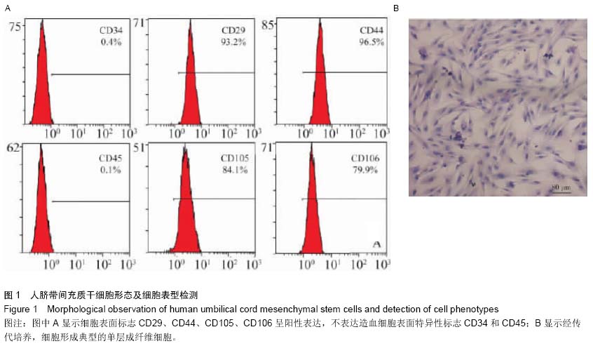
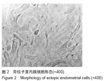
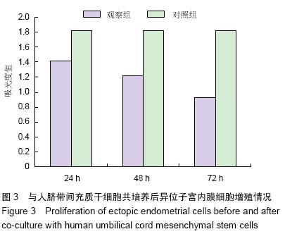
| [1] 徐丽南,徐冰南,陈淑琴,等.人脐带间充质干细胞影响子宫内膜异位症细胞的增殖及凋亡[J].中国组织工程研究,2012,16(10): 1765-1768.
[2] 杨晓清,徐云钊,虞琪,等.人脐带间充质干细胞对大鼠受损子宫内膜的影响[J].江苏医药,2013,39(10):1130-1133.
[3] 张玉泉,杨晓清.间充质干细胞与妇产科[J].南通大学学报:医学版, 2012,32(1):1-6.
[4] 周长辉.人脐带Wharton's jelly及子宫内膜间充质干细胞免疫调节作用研究[D]. 郑州:郑州大学,2010.
[5] Ali IU. Gatekeeper for endometrium: the PTEN tumor suppressor gene. J Natl Cancer Inst. 2000;92(11):861-863.
[6] 臧传宝,李栋.间充质干细胞向上皮细胞的诱导分化及应用研究进展[J].国际儿科学杂志,2011,38(6):616-619.
[7] Yang S, Sun HM, Yan JH, et al. Conditioned medium from human amniotic epithelial cells may induce the differentiation of human umbilical cord blood mesenchymal stem cells into dopaminergic neuron-like cells. J Neurosci Res. 2013;91(7): 978-986.
[8] 张真真,桂涛,成臣,等.人子宫内膜间充质干细胞的分离、培养与鉴定[J].江苏医药,2014,40(9):1000-1002.
[9] 杨新园,乔杰,郑鹏生,等.子宫内膜干细胞研究进展[J].现代妇产科进展,2008,17(7):546-548.
[10] 袁华,王沂峰,赵华,等.骨髓间充质干细胞对小鼠异位子宫内膜的作用观察[J].山东医药,2012,52(46):40-41.
[11] 司宇.经血源性子宫内膜间充质干细胞向平滑肌细胞诱导分化的初步研究[D]. 哈尔滨:哈尔滨医科大学,2014.
[12] 彭艳.经血源性子宫内膜间充质干细胞体外分离培养及初步鉴定[D]. 哈尔滨:哈尔滨医科大学,2012.
[13] 丛青.小鼠骨髓间充质干细胞向子宫内膜腺上皮分化的蛋白质组学研究[D].上海:复旦大学,2012.
[14] 路平,赵潇丹,郝玉娟,等.小鼠骨髓间充质干细胞在子宫内膜损伤小鼠子宫内膜中的定位[J].郑州大学学报:医学版,2015,50(1): 101-103,104.
[15] 张雯碧,程明军,徐丛剑,等.小鼠骨髓间充质干细胞向子宫内膜上皮细胞方向分化的体外实验[J].现代妇产科进展,2010,19(4): 257-260.
[16] Gebel HM, Braun DP, Tambur A, et al. Spontaneous apoptosis of endometrial tissue is impaired in women with endometriosis. Fertil Steril. 1998;69(6):1042-1047.
[17] Simões IN, Boura JS, dos Santos F, et al. Human mesenchymal stem cells from the umbilical cord matrix: successful isolation and ex vivo expansion using serum-/ xeno-free culture media. Biotechnol J. 2013;8(4): 448-458.
[18] Esposito M, Lucariello A, Costanzo C, et al. Differentiation of human umbilical cord-derived mesenchymal stem cells, WJ-MSCs, into chondrogenic cells in the presence of pulsed electromagnetic fields. In Vivo. 2013;27(4):495-500.
[19] 王永刚.大鼠骨髓间充质干细胞向子宫内膜细胞分化的体外实验[D].长沙:中南大学,2012.
[20] 赵静.骨髓间充质干细胞移植治疗薄型子宫内膜的实验研究[D]. 长沙:中南大学,2013.
[21] 陈洪流,陈振勇.Survivin和第10号染色体缺失的磷酸酶及张力蛋白同源物基因基因在肝癌组织中的表达及其相关性[J].中华实验外科杂志,2013,30(6):1283-1285.
[22] 雷莹,陈杭,许红,等.PTEN基因的沉默对骨髓间充质干细胞体外生物学特性和体内移植治疗心肌梗死的影响[J].临床心血管病杂志,2011,27(4):307-312.
[23] Roszek K, Bomastek K, Dro?d?al M, et al. Dramatic differences in activity of purines metabolizing ecto-enzymes between mesenchymal stem cells isolated from human umbilical cord blood and umbilical cord tissue. Biochem Cell Biol. 2013;91(6):519-525.
[24] 戴磊.间充质干细胞-抗肿瘤基因复合物治疗肿瘤的研究进展[J].肿瘤研究与临床,2013,25(11):788-791.
[25] 陈毅. MMP-2和PTEN双基因干预对骨髓间充质干细胞移植治疗脊髓损伤的影响[D].沈阳:中国医科大学,2011.
[26] 林叔陈,张凤春,张雁云,等.肿瘤干细胞的调控机制研究进展[J].现代肿瘤医学,2009,17(1):183-186.
[27] Yun SP, Lee SJ, Oh SY, et al. Reactive oxygen species induce MMP12-dependent degradation of collagen 5 and fibronectin to promote the motility of human umbilical cord-derived mesenchymal stem cells. Br J Pharmacol. 2014;171(13):3283-3297.
[28] 高磊,潘铁军,武国军,等.第10号染色体缺失的磷酸酶及张力蛋白同源物基因对人前列腺癌细胞株PC-3转移及基质金属蛋白酶-2、基质金属蛋白酶-9表达的影响[J].中华实验外科杂志,2013, 30(12): 2490-2492.
[29] 闫旭,阚世廉,金晔,等.第10号染色体缺失的磷酸酶及张力蛋白同源物基因/雷帕霉素靶蛋白调节细胞骨架构建在神经细胞分化过程中的变化及意义[J].中华实验外科杂志,2013,30(7):1387- 1389.
[30] Yang S, Xue DD, Wu B, et al. Pleiotrophin is involved in the amniotic epithelial cell-induced differentiation of human umbilical cord blood-derived mesenchymal stem cells into dopaminergic neuron-like cells. Neurosci Lett. 2013;539: 86-91.
[31] Bai H, Chen P, Tang GQ, et al. Relations between reactive oxygen species and Raman spectral variations of human umbilical cord mesenchymal stem cells with different viability. Laser Physics. 2011;21(6):1122-1129.
[32] Harada T, Kaponis A, Iwabe T, et al. Apoptosis in human endometrium and endometriosis. Hum Reprod Update. 2004;10(1):29-38.
[33] Mutter GL, Lin MC, Fitzgerald JT, et al. Changes in endometrial PTEN expression throughout the human menstrual cycle. J Clin Endocrinol Metab. 2000;85(6):2334- 2338.
[34] 陈毅,尹西盟,董宝铁,等.PTEN基因沉默对BMSC氧糖剥夺模型的保护作用及其对Bcl-2凋亡通路的影响[J].中国医科大学学报,2011,40(7):600-604. |
lb.jpg)