中国组织工程研究 ›› 2015, Vol. 19 ›› Issue (36): 5746-5752.doi: 10.3969/j.issn.2095-4344.2015.36.002

• 骨髓干细胞 bone marrow stem cells • 上一篇    下一篇

肝细胞生长因子基因转染缺氧培养骨髓间充质干细胞凋亡相关蛋白的影响

蔡文钦1,王隽苼1,苏津自1,江金峰1,姚月娴2   

  1. 1福建医科大学附属第一医院心内科,高血压研究所,福建省福州市  350005; 2福建省立医院北院福建省老年医院心内科,福建省福州市  350003
  • 出版日期:2015-09-03 发布日期:2015-09-03
  • 通讯作者: 姚月娴,硕士,主治医师,福建省立医院北院福建省老年医院心内科,福建省福州市 350003
  • 作者简介:蔡文钦,男,1980年生,福建省龙岩市人,汉族,2010年福建医科大学毕业,硕士,主治医师,主要从事对比剂肾病及干细胞研究。
  • 基金资助:

    福建省卫生厅青年基金(2011-1-32,2011-1-15);福建省自然科学基金(2014J01433)

Expressions of apoptosis-related proteins in hepatocyte growth factor-transfected bone marrow mesenchymal stem cells under hypoxia conditions 

Cai Wen-qin1, Wang Jun-sheng1, Su Jin-zi1, Jiang Jin-feng1, Yao Yue-xian2   

  1. 1Department of Cardiology, Fujian Institute of Hypertension Research, First Affiliated Hospital, Fujian Medical University, Fuzhou 350005, Fujian Province, China; 2Department of Cardiology, Fujian Provincial Geriatric Hospital, Fuzhou 350003, Fujian Province, China
  • Online:2015-09-03 Published:2015-09-03
  • Contact: 姚月娴,硕士,主治医师,福建省立医院北院福建省老年医院心内科,福建省福州市 350003
  • About author:Cai Wen-qin, Master, Attending physician, Department of Cardiology, Fujian Institute of Hypertension Research, First Affiliated Hospital, Fujian Medical University, Fuzhou 350005, Fujian Province, China
  • Supported by:

    the Youth Fund of Fujian Provincial Department of Health, No. 2011-1-32, 2011-1-15; the Natural Science Foundation of Fujian Province, No. 2014J01433

摘要:

背景:研究已证实肝细胞生长因子基因转染可提高骨髓间充质干细胞移植效果,但其具体机制尚未完全明确。

目的:探讨肝细胞生长因子基因转染对缺氧、无血清培养骨髓间充质干细胞c-Met、Bax、Bcl-2、Caspase-3表达及细胞迁移的影响。

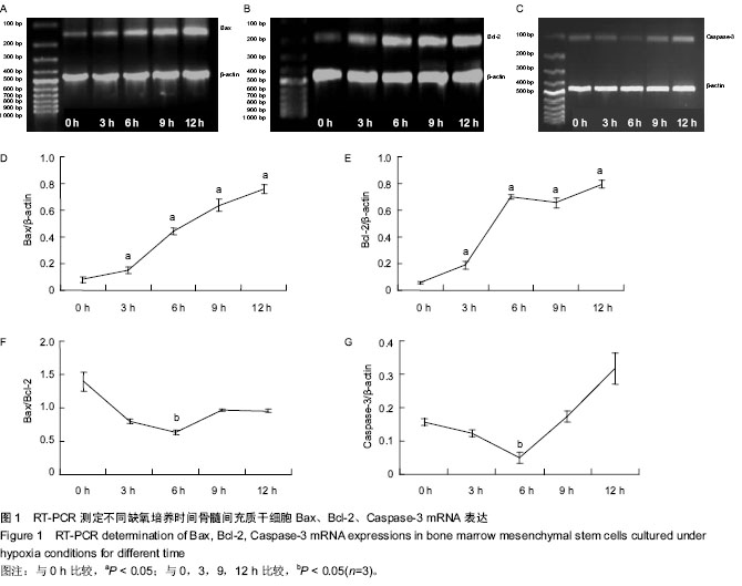
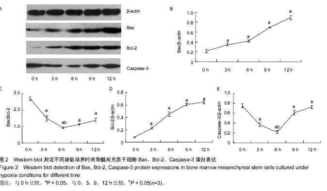
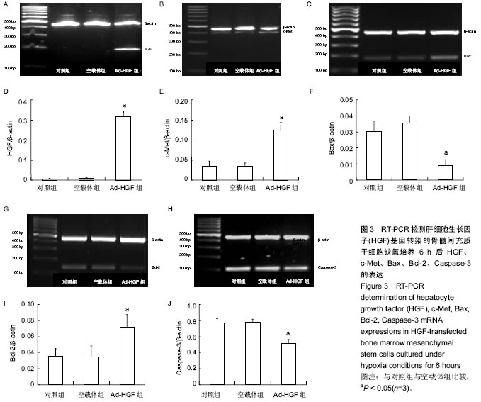
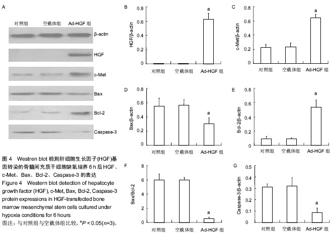
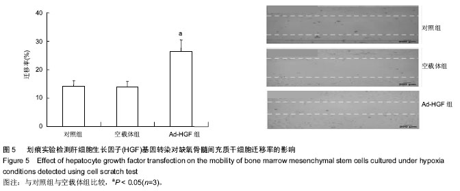
方法:①贴壁法体外分离、扩增培养骨髓间充质干细胞。用x-gal染色法检测含肝细胞生长因子基因的重组腺病毒对骨髓间充质干细胞的感染率。②在缺氧、无血清条件下培养0,3,6,9,12 h,采用RT-PCR及Western blot测定骨髓间充质干细胞Bax、Bcl-2、Caspase-3的表达。③骨髓间充质干细胞缺氧、无血清培养6 h,采用RT-PCR及Western blot测定HGF、c-Met、Bax、Bcl-2、Caspase-3的表达。④细胞迁移划痕法观察缺氧、无血清培养6 h后肝细胞生长因子转染对骨髓间充质干细胞迁移的影响。

结果与结论:①重组腺病毒对骨髓间充质干细胞的转染率与病毒感染复数具有量效关系,病毒感染复数为150时细胞的感染率达96.4%。②随着缺氧时间的延长骨髓间充质干细胞Bax、Bcl-2表达逐渐升高(P < 0.05),缺氧6 h时Bax/Bcl-2比值、Caspase-3蛋白达到最小(P < 0.05)。③缺氧及无血清培养6 h后,与对照组及空载体组相比,Ad-HGF组HGF、c-Met、Bcl-2表达增高,而Bax、Caspase-3表达下降(P < 0.05),对照组与空载体组差异无显著性意义(P > 0.05)。④Ad-HGF组骨髓间充质干细胞在缺氧培养6 h后,迁移率比对照组及空载体组高(P < 0.05)。结果表明肝细胞生长因子基因转染上调缺氧、无血清培养骨髓间充质干细胞c-Met、Bcl-2表达,下调Bax、Caspase-3表达,增强骨髓间充质干细胞迁移能力。

 中国组织工程研究杂志出版内容重点:干细胞;骨髓干细胞;造血干细胞;脂肪干细胞;肿瘤干细胞;胚胎干细胞;脐带脐血干细胞;干细胞诱导;干细胞分化;组织工程

关键词: 干细胞, 骨髓干细胞, 肝细胞生长因子, 骨髓间充质干细胞, 凋亡蛋白, 缺氧培养, 细胞迁移, 福建省自然科学基金

Abstract:

BACKGROUND: Previous studies have demonstrated that hepatocyte growth factor (HGF) gene transfection can improve the effectiveness of bone marrow mesenchymal stem cell transplantation, but the mechanism is still unclear.

OBJECTIVE: To observe the effects of HGF gene transfection on c-MET, Bax, Bcl-2, Caspase-3 of bone marrow mesenchymal stem cells cultured under hypoxia and serum-free conditions.

METHODS: (1) Bone marrow mesenchymal stem cells were isolated and amplified in vitro by differential adhesion method. The infection efficiency of recombinant adenovirus Ad-HGF in bone marrow mesechymal stem 
cells was tested by x-gal staining. (2) Bone marrow mesenchymal stem cells were cultured under hypoxia and serum-free conditions for 0, 3, 6, 9, 12 hours. RT-PCR and western blot assays were used to evaluate the expression of Bax, Bcl-2, Caspase-3. (3) Bone marrow mesenchymal stem cells were cultured under hypoxia and serum-free conditions for 6 hours, and RT-PCR and western blot assays were adopted to detect HGF, c-Met, Bax, Bcl-2 and Caspase-3. (4) Cell scratch test was used to detect the effect of HGF transfection on the migration of bone marrow mesenchymal stem cells cultured under hypoxia and serum-free conditions for 6 hours.

RESULTS AND CONCLUSION: (1) Transfection efficiency of bone marrow mesenchymal stem cells was increased with multiplicity of infection in a dose-dependent manner. When the multiplicity of infection was 150, the transfection efficiency was 96.4%. (2) Expressions of Bax and Bcl-2 were gradually increased with hypoxia time (P < 0.05). The Bax/Bcl-2 ratio and Caspase-3 expression reach the minimum at 6 hours of hypoxia (P < 0.05). (3) Compared with the control and Ad-LacZ groups, the expressions of HGF, c-Met, Bcl-2 increased, and the expressions of Bax and Caspase-3 decreased in the Ad-HGF group after 6 hours of culture under hypoxia and serum-free conditions (P < 0.05). There was no significant difference between the control and Ad-LacZ groups. (4) The mobility of bone marrow mesenchymal stem cells was higher in the Ad-HGF group than the control group and Ad-LacZ groups after 6 hours of culture under hypoxia (P < 0.05). These findings indicate that transfection of HGF in bone marrow mesenchymal stem cells can increase the expression of c-Met, Bcl-2 and decrease the expression of Bax, Caspase-3 under hypoxia and serum-free conditions, which also enhance the mobility of bone marrow mesenchymal stem cells under hypoxia and serum-free conditions.

 中国组织工程研究杂志出版内容重点:干细胞;骨髓干细胞;造血干细胞;脂肪干细胞;肿瘤干细胞;胚胎干细胞;脐带脐血干细胞;干细胞诱导;干细胞分化;组织工程

Key words: Bone Marrow, Mesenchymal Stem Cells, Hepatocyte Growth Factor, Cell Hypoxia

中图分类号: