1 软骨退行性改变 Cartilage degeneration
常见误区:①膝关节间隙发现无回声区即考虑为关节积液。②关节软骨变薄可诊断为软骨退行性改变。
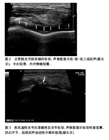
正常膝关节软骨在高频超声上呈覆盖于骨质表面的“高-低-高”三层结构(图2),表面高回声轮廓线是由软骨-滑液腔界面形成,软骨下骨界面则形成了深面高回声线,而软骨则呈低-无回声,易被初学者诊断为关节积液。正常膝关节软骨中央较厚,向边缘逐渐变薄,移行自然[1]。超声主要从软骨表面形态、软骨内回声以及厚度改变来诊断软骨退行性改变(退变),而不能仅根据软骨变薄来诊断退变。相对于测量软骨厚度而言,软骨表面是否光滑以及其内透声是否清晰更能提示软骨的早期异常改变。
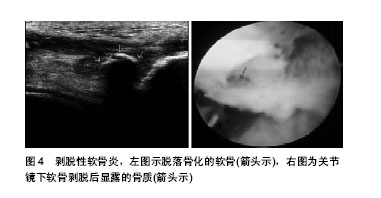
国际软骨修复协会( International Cartilage Repair Society,ICRS) 提出软骨分级标准为0-4级,0级: 正常软骨;1级: 接近正常,表面凹陷(1A),表面裂隙(1B);2级: 病变范围小于软骨厚度的1 /2;3级: 软骨缺损超过软骨厚度的1/2;4级: 全层软骨缺损,病变腐蚀软骨下骨板[2]。许多疾病均会引起软骨退变,如类风湿性关节炎(图3)、骨性关节炎等,当软骨病变严重脱落时,常导致软骨下骨质暴露、受损(图4)。但软骨退变早期发生软骨基质丢失或深层纤维断裂软骨变软等细微改变,此时超声常无法捕捉其变化,表现为正常软骨回声,易做出假阴性诊断。由于膝关节腔空间狭小,其内组织结构复杂,当膝关节发生炎性改变时,增生的滑膜、纤维渗出的覆盖,可使软骨回声显示不清,此时易将正常软骨误诊为病变软骨组织。
2 滑膜钙化 Synovial calcification
常见误区:①滑膜病变内出现钙化,即可诊断为恶性肿瘤。②钙化在滑膜良性和恶性病变中均可出现,对超声诊断无太大价值。
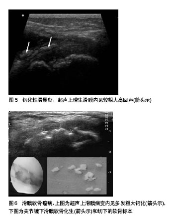
超声对钙化具有高度的敏感性,对强回声的钙化或骨化灶及关节游离体具有独特的诊断优势,常作为诊断疾病的重要依据。膝关节滑膜病变内钙化可分为斑块状粗大钙化和颗粒状小钙化两种形态。斑块状粗大钙化声像图上呈斑块状强回声,形态不规则,后方多伴声影,常见于滑膜软骨瘤病及钙化性滑囊炎(图5)。微小钙化声像图上呈颗粒状、点状强回声,可呈簇状聚集或散在分布,后方一般无声影,可见于恶性肿瘤(如滑膜肉瘤)和良性病变(如滑膜软骨瘤病),因此,对于小钙化的鉴别是判断良恶性的关键所在。
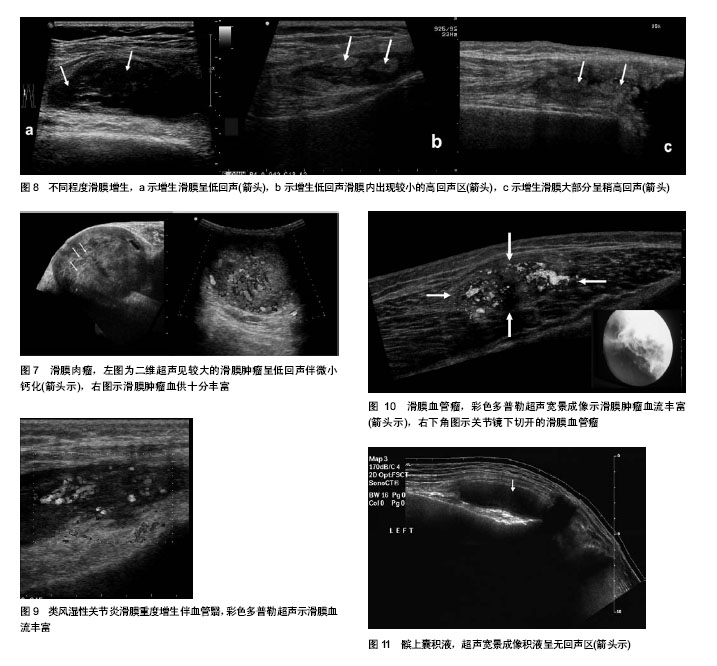
滑膜软骨瘤病(图6)为临床罕见的一种常累及单侧滑膜组织的疾病,其主要病理学特征是在滑膜组织内出现骨或软骨组织的小结节[3-4]。早期,软骨结节未形成钙化或骨化,也未脱落形成关节游离体时,超声表现为低回声团块,易被误诊为色素沉着绒毛结节性滑膜炎。当软骨小体形成后,超声上即出现颗粒样钙化,此时与滑膜肉瘤(图7)的微钙化很难鉴别,需要参考其他声像图特征。当软骨小体进一步骨化连接成片时,超声上表现为斑片状粗大钙化,如发生在滑膜内此病不难诊断,但注意与慢性肌腱病中的钙化相鉴别,后者钙化发生在增厚的肌腱内。
3 滑膜病变回声强度 Echo intensity from synovial lesions
常见误区:①滑膜恶性团块多为低回声,良性团块多为中、高回声。②弥漫增厚滑膜呈低回声表示炎症明显,呈高回声表示炎症消退。
膝关节滑膜是一层血管高度丰富的结缔组织膜,由内膜和滑膜下组织组成。滑膜内膜是一层由1-4层连接松散的细胞构成的滑膜衬里[5]。滑膜细胞由A细胞(巨噬细胞样细胞)和B细胞(成纤维细胞样细胞)构成,A细胞的主要功能是吞噬进入关节的内源性或外源性异物,是各种膝关节疾病出现滑膜反应的主要原因。滑膜B细胞则与关节内透明质酸-蛋白质的合成分泌有关[6],透明质酸参与滑液的构成,具有润滑关节的作用。滑膜下组织的结构随部位不同而变化,可为纤维性的、网状的、纤维网状的、脂肪网状的或脂肪组织构成,内含大量血管、淋巴管[7-9]。
在滑膜疾病的早期,滑膜下结缔组织的增生并不显著,故在超声上表现为略低回声的增厚滑膜(图8a)。当病情进展,滑膜受到反复炎症刺激后,可导致滑膜及滑膜下结缔组织纤维增生,超声上表现为增厚滑膜低回声内出现较小的高回声区(图8b),甚至表现为增生滑膜大部分呈中高回声(图8c),高回声区的出现常提示长期慢性滑膜炎。
增厚的滑膜,其内部回声与炎性因子所致复杂的滑膜病理改变有关。临床上增厚滑膜内部回声较均匀者常提示良性滑膜疾病。在滑膜肉瘤的超声表现中,较大的肿瘤由于瘤体的增长快于瘤体内血管的增生,瘤体内常发生局灶性坏死,在超声上常表现为局部低回声或无回声区,故较大肿瘤则常常表现为边界不规则的分叶状肿块,内部回声不均匀。但较小的肿瘤,一般表现为边界较为清晰的结节状类圆形,其内部多为较均匀实性低回声区[10]。
4 滑膜血流情况 Blood flow in the synovium
常见误区:①滑膜局灶性病变内血流越丰富,提示恶性肿瘤可能性越大。②弥漫增生的滑膜组织内血流丰富,提示炎症;如未探及血流信号,提示无炎症。
超声对于血流十分敏感,能够有效的分辨出组织内的血流信号。在受到炎症刺激后,滑膜下组织内的血管成分可因炎性因子的刺激而增生,如类风湿性关节炎中滑膜明显增生伴大量血管翳形成(图9)。大多数滑膜恶性肿瘤,由于其生长所需,及其独特的病理变化,瘤体内部血流多较为丰富。但在一些良性滑膜病变中,亦可见到不同程度增生的滑膜下血管组织。滑膜血管瘤(图10)是一种少见的良性血管疾病,起源于滑膜下层的间充质[11]。其病理表现为关节内局限性或弥慢性肿物,为海绵状的增生血管组织,呈分支状。在超声上病变表现为低至中强回声的软组织肿块,有时可见丰富血流信号[12] 。
值得注意的是,随着慢性病程的进展,滑膜下组织内的纤维间质成分也逐渐增多。至病程中晚期,虽然滑膜内的血流依然较正常为多,但在超声图像上由于受到间质成分的影响,弥漫增生的滑膜内血流信号不易被探及,但此时炎症依然存在。
5 滑囊积液 Synovial effusion
常见误区:①所有的滑膜积液均由滑膜疾病所导致。②膝关节滑囊积液均呈无回声区。③髌上囊内出现低或无回声区一定是积液。④超声未探及滑膜或关节腔积液时,即可排除滑膜炎症。⑤滑囊积液内如出现低回声团块,系增生的滑膜组织。
滑膜积液是滑膜组织对损伤的炎症性反应。滑膜损伤后,局部充血、水肿、白细胞及纤维素渗出,当渗出速度超过代偿性吸收速度时,积液形成。临床上滑膜积液成分的分析对于疾病的诊断有着十分重要的意义[13-14]。超声是诊断膝关节积液的最佳方法,声像图上呈无或低回声区(图11)。值得注意的是,在生理状态下膝关节很多滑囊仅是一个潜在的腔隙,在正常情况下,目前的超声及其他影像的分辨率还不能显示这些滑囊(如髌前皮下囊、髌下浅囊、鹅足腱下滑囊等)。当超声检查发现这些部位滑囊液体时,多可认为是病理情况。然而,也有一些滑囊在生理状态下即含有极少量滑液,与周围组织形成很好的界面回声,高频超声可清晰显示这些滑囊的结构形态,为薄层的片状无回声区(如髌上囊等),这些部位在生理状态下超声可见深1.0-2.0 mm的极少量积液,超过2 mm深的积液则被视为病理情况[15]。
关节积液常常为非特异性,并不一定都表示滑膜炎症。大多数膝关节滑膜病变均合并髌上囊或关节腔积液。骨关节炎,关节急慢性损伤、感染、关节置换术后、全身性疾病(如风湿病、痛风)等,均可出现关节积液。当髌上囊内滑膜弥漫性增生呈较低回声时,声像图上酷似积液(图12)。增大仪器增益常能显示增生的滑膜组织,另外,通过挤压扫查可鉴别积液与低回声的增生滑膜。
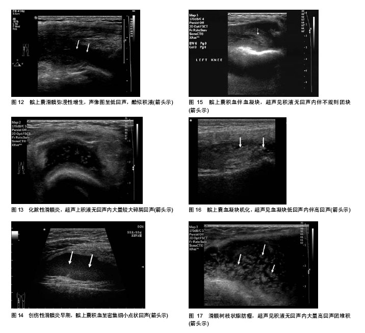
滑囊积液伴有其他回声,对滑膜病变具有重要的提示价值。①积液内出现细小碎屑回声,探头加压,可见其缓慢移动,常代表滑膜组织碎片。②积液内出现较大碎屑样回声,探头加压可见移动,如患者有发热病史,应考虑化脓性关节炎(图13)。③创伤性滑膜炎早期,滑囊内大量新鲜积血时,声像图表现为滑囊内充满密集细小点状回声(图14)。积液内出现较低回声、不规则团块,则应考虑血凝块回声(图15),如血凝块机化时,团块将出现高低混合性回声或高回声(图16)。④积液内出现高回声团堆积呈绒毛状突起,探头加压不移动,但可被压缩,可见于一种罕见病变-滑膜树枝状脂肪瘤(图17),此高回声团系滑膜下脂肪浸润引起,CT或MRI检查时可显示病变内的脂肪成分,则更有助于确诊。
临床上往往将滑膜积液的出现作为诊断滑膜病变的首要征象。但值得注意的是,当滑膜或关节腔积液量很少时,超声不能获得良好的滑膜与积液的对比界面,从而超声检查常常不易发现。此时,易误将病变滑膜当成正常滑膜。
6 累及膝关节内其他结构 Lesions involving intraarticular structures
常见误区:当滑膜病变累及膝关节内其他结构时,提示为恶性滑膜疾病。
临床常见的恶性滑膜病变滑膜肉瘤是一类恶性程度较高的软组织肿瘤,好发于四肢近关节旁,其组织来源是由未分化间叶细胞发生的具有滑膜分化特点的恶性肿瘤。在超声上多表现为圆形或分叶状肿块,其内回声多与肿物病理组成相关,可呈低回声区、不均匀回声区等。当其肿瘤侵犯关节腔内其他组织时,可有相应超声图像显示[16]。但在一些良性滑膜病变中,弥漫增生的滑膜也常常累及关节内的其他正常结构。类风湿性关节炎为一种主要侵犯关节滑膜的疾患,在类风湿性关节炎晚期(慢性纤维化期)时,滑膜增生显著,甚至可以形成乳头状突起,逐渐向关节周围及软骨边缘部扩展。同时,于关节软骨边缘的滑膜反折处,增生的富于血管翳样肉芽组织向周围软骨扩展蔓延,从而渐进性破坏并取代软骨组织,非常类似于局限性侵袭生长的肿瘤特征[17]。弥漫型色素绒毛结节性滑膜炎(PVNS),随着长期慢性炎症刺激,其弥漫增生的滑膜常可侵及关节软骨及半月板[18],但其较少出现溶骨性的骨质破坏区域,弥漫增生的滑膜内出现钙化也较滑膜肉瘤少见。弥漫型滑膜血管瘤其病变界限多不清楚,其肿瘤组织也可出现侵犯关节囊周围的韧带和肌层等组织的特征。
.jpg)