中国组织工程研究 ›› 2014, Vol. 18 ›› Issue (45): 7246-7249.doi: 10.3969/j.issn.2095-4344.2014.45.006

• 骨髓干细胞 bone marrow stem cells • 上一篇    下一篇

优化培养基加快骨髓间充质干细胞的诱导分化

李 倩1,杨 军2   

  1. 1重庆市第五人民医院神经内科,重庆市 400062;2重庆医科大学附属第一医院神经内科,重庆市 400016
  • 出版日期:2014-11-05 发布日期:2014-11-05
  • 通讯作者: 杨军,重庆医科大学附属第一医院神经内科,重庆市 400016
  • 作者简介:李倩,女,1984年生,硕士,主要从事脑血管疾病的基础与临床研究。
  • 基金资助:

    重庆市卫生局科学基金(09-2-335);重庆市自然科学基金(CSTC,2005BB5315)

Optimized medium accelerates differentiation of bone marrow mesenchymal stem cells

Li Qian1, Yang Jun2   

  1. 1Department of Neurology, the Fifth People’s Hospital of Chongqing, Chongqng   400062, China; 2Department of Neurology, the First Affiliated Hospital, Chongqing Medical University, Chongqing 400016, China
  • Online:2014-11-05 Published:2014-11-05
  • Contact: Yang Jun, Department of Neurology, the First Affiliated Hospital, Chongqing Medical University, Chongqing 400016, China
  • About author:Li Qian, Master, Department of Neurology, the Fifth People’s Hospital of Chongqing, Chongqng 400062, China
  • Supported by:

    the Science Foundation of Chongqing Health Bureau, No. 09-2-335; the Natural Science Foundation of Chongqing, No. CSTC, 2005BB5315

摘要:

背景:研究发现骨髓间充质干细胞在一定的诱导条件下能分化成神经元、神经胶质等神经细胞,这一定程度上解决了种子细胞来源的难题。目前采用的诱导方法各有不同,所诱导分化的效率也不尽相同。


目的:观察不同抗氧化剂体外诱导大鼠骨髓间充质干细胞向神经样细胞分化的效果。


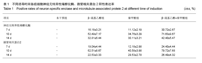
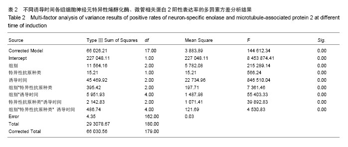
方法:将Wistar大鼠骨髓间充质干细胞分为4组,使用不同神经细胞诱导剂诱导分化,分别为未干预组、β-巯基乙醇组、维甲酸组、β-巯基乙醇联合维甲酸组,诱导5 h,12 h,1 d,3 d,5 d,7 d,10 d后,分别观察细胞形态变化及检测神经元细胞特异性抗原神经元特异性烯醇化酶、微管相关蛋白2阳性率表达差异。


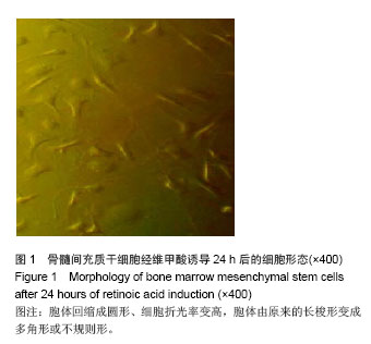
结果与结论:未干预组骨髓间充质干细胞形态无明显改变,其他3组细胞逐渐变成纺锤形,并生出多个小突起,互相连接成网,呈现神经元样细胞形态;免疫细胞化学染色显示β-巯基乙醇联合维甲酸组在诱导10 d后其效率最高,神经元特异性烯醇化酶、微管相关蛋白2表达阳性率分别是71.63%和79.72%。结果显示β-巯基乙醇联合维甲酸可加快诱导骨髓间充质干细胞向神经元样细胞分化。


中国组织工程研究杂志出版内容重点:干细胞;骨髓干细胞;造血干细胞;脂肪干细胞;肿瘤干细胞;胚胎干细胞;脐带脐血干细胞;干细胞诱导;干细胞分化;组织工程


全文链接:

关键词: 干细胞, 骨髓干细胞, 诱导剂, 骨髓间充质干细胞, 诱导分化, 维甲酸, β-巯基乙醇, 神经元特异性烯醇化酶, 微管相关蛋白2, 重庆市自然科学基金

Abstract:

BACKGROUND: Studies have found that bone marrow mesenchymal stem cells, under certain conditions, can be induced to differentiate into neurons and glial cells, which to some extent solves the problem of the source of seed cells. Induction methods currently used are different, and their efficiencies are not the same.

 
OBJECTIVE: To observe the effects of different antioxidants on differentiation of rat bone marrow mesenchymal stem cells into neuron-like cells in vitro.
METHODS: Bone marrow mesenchymal stem cells from Wistar rats were divided into four groups: non-intervention group, β-mercaptoethanol group, retinoic acid group, β-mercaptoethanol+retinoic acid group. Changes in cell morphology and positive rate of neuron-specific enolase and microtubule-associated protein 2 were observed and detected at 5 hours, 12 hours, 1 day, 3 days, 5 days, 7 days, and 10 days after induction.
RESULTS AND CONCLUSION: Except non-intervention group, bone marrow mesenchymal stem cells in the other three groups were gradually becoming spindle-shaped, and gave birth to many small protrusions that were interconnected into a network, showing neuron-like cell morphology. Immunocytochemical staining showed that the efficiency of the β-mercaptoethanol+retinoic acid group was the highest at 10 days after induction, and the positive rates of neuron-specific enolase and microtubule-associated protein 2 were 71.63% and 79.72%, respectively. The results show that β-mercaptoethanol can be combined with retinoic acid to accelerate the differentiation of bone marrow mesenchymal stem cells into neuron-like cells.

中国组织工程研究杂志出版内容重点:干细胞;骨髓干细胞;造血干细胞;脂肪干细胞;肿瘤干细胞;胚胎干细胞;脐带脐血干细胞;干细胞诱导;干细胞分化;组织工程


全文链接:

Key words: bone marrow, mesenchymal stem cells, cell differentiation, tretinoin, mercaptoethanol

中图分类号: