中国组织工程研究 ›› 2014, Vol. 18 ›› Issue (37): 6019-6023.doi: 10.3969/j.issn.2095-4344.2014.37.021

• 干细胞与中医药 stem cells and traditional Chinese medicine • 上一篇    下一篇

红景天苷诱导骨髓间充质干细胞向神经元样细胞分化中的Ca2+/CaM信号通路

赵  玲1,赵红斌2,潘  茜2,李  根2,王九娜2,唐俊杰2   

  1. 1甘肃中医学院,甘肃省兰州市  730000
    2解放军兰州军区兰州总医院,甘肃省兰州市  730000
  • 修回日期:2014-08-09 出版日期:2014-09-03 发布日期:2014-09-03
  • 通讯作者: 赵红斌,博士,硕士生导师,主任技师,解放军兰州军区兰州总医院,甘肃省兰州市 730000
  • 作者简介:赵玲,女,1988年生,甘肃省武威市人,汉族,甘肃中医学院在读硕士,主要从事中药与干细胞方面的研究。
  • 基金资助:

    国家自然科学基金面上项目资助(81073156),课题名称:红景天苷诱导间充质干细胞向神经细胞分化的作用及分子机制

Salidroside induces the differentiation of mouse bone marrow mesenchymal stem cells into neuron-like cells mediated by calcium/calmodulin signaling pathway

Zhao Ling1, Zhao Hong-bin2, Pan Qian2, Li Gen2, Wang Jiu-na2, Tang Jun-jie2   

  1. 1Gansu College of Traditional Chinese Medicine, Lanzhou 730000, Gansu Province, China
    2Institute of Orthopaedics, Lanzhou General Hospital of Lanzhou Military Region, Lanzhou 730000, Gansu Province, China
  • Revised:2014-08-09 Online:2014-09-03 Published:2014-09-03
  • Contact: Zhao Hong-bin, M.D., Master’s supervisor, Chief technician, Institute of Orthopaedics, Lanzhou General Hospital of Lanzhou Military Region, Lanzhou 730000, Gansu Province, China
  • About author:Zhao Ling, Studying for master’s degree, Gansu College of Traditional Chinese Medicine, Lanzhou 730000, Gansu Province, China
  • Supported by:

    the National Natural Science Foundation of China, No. 81073156

摘要:

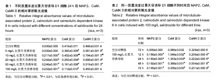
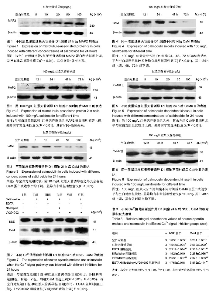
背景:课题组前期研究表明,红景天苷能诱导骨髓间充质干细胞向神经元样细胞定向分化,Ca2+信号是实现其生物学信号传导的重要途径之一
目的:探讨Ca2+/CaM 信号通路在红景天苷诱导骨髓间充质干细胞向神经细胞定向分化中的作用及机制。
方法:实验分为空白对照组和红景天苷诱导组,红景天苷诱导组将不同质量浓度红景天苷(5,10,20,50,100 mg/L)作用骨髓间充质干细胞24 h和100 mg/L红景天苷作用骨髓间充质干细胞12,24,48,72 h。采用Western blot方法分别检测红景天苷诱导骨髓间充质干细胞后神经标志分子MAP2和Ca2+/CaM信号通路中关键蛋白CaM、CaMKⅡ的表达水平。另外实验设阻断剂组,分别加Ca2+信号通路特异性阻断剂:500 µmol/L EGTA (细胞外Ca2+螯合剂)、1 mmol/L Nifedipine (L型Ca2+通道阻断剂)、10 mmol/L LY294002 (PI3K抑制剂)分别作用细胞30 min后,再加入100 mg/L红景天苷作用细胞24 h,采用Western blot方法检测阻断Ca2+/CaM信号通路后NSE、CaM的表达情况。
结果与结论:①红景天苷诱导后,MAP2的表达上调(P < 0.01),说明红景天苷可诱导骨髓间充质干细胞分化为神经细胞。②不同质量浓度红景天苷诱导骨髓间充质干细胞24 h后,10 mg/L红景天苷组CaM、CaMKⅡ的表达与其他诱导组比较显著上调(P < 0.01);同一质量浓度红景天苷诱导72 h后CaM、CaMKⅡ表达明显下调(P < 0.01)。③阻断细胞外Ca2+和PI3K信号通路后,NSE与CaM的表达水平较红景天苷诱导组上调(P < 0.05)。结果表明,红景天苷通过抑制Ca2+/CaM信号通路实现骨髓间充质干细胞向神经细胞定向分化。


中国组织工程研究杂志出版内容重点:干细胞;骨髓干细胞;造血干细胞;脂肪干细胞;肿瘤干细胞;胚胎干细胞;脐带脐血干细胞;干细胞诱导;干细胞分化;组织工程


全文链接:

关键词: 干细胞, 骨髓干细胞, 红景天苷, 骨髓间充质干细胞, Ca2+/CaM信号通路, 神经细胞, 国家自然科学基金

Abstract:

BACKGROUND: Our previous studies have shown that salidroside can induce bone marrow mesenchymal stem cells directly into neuron-like cells, and Ca2+ signal is one important way to achieve its biological signal transduction.
OBJECTIVE: To investigate the role and mechanism of the calcium/calmodulin (Ca2+/CaM) signaling pathway inducing bone marrow mesenchymal stem cells to differentiate into nerve cells.
METHODS: Bone marrow mesenchymal stem cells were divided into two groups: control groups and salidroside groups. Salidroside groups were induced with different concentrations of salidroside (5, 10, 20, 50 and 100 mg/L) for 24 hours and 100 mg/L salidroside was added to culture cells for different time (12, 24, 48 and 72 hours). Western blot assay was used to detect the expression levels of neural cell marker, microtubule-associated protein 2, and the important protein of Ca2+/CaM signaling pathway: CaM and calmodulin dependent kinase II (CaMK II). Then Ca2+/CaM signaling pathway specific blockers were applied to cells respectively for 30 minutes, including 500 µmol/L EGTA (Ca2+ chelator), 1 mmol/L Nifedipine(L-type Ca2+ channel blocker) and 10 mmol/L LY294002 (PI3K inhibitor). Then, 100 mg/L salidroside was added and cultured for 24 hours. Western blot assay was used to detect the expression of neuron-specific enolase and CaM in the Ca2+/CaM signaling pathway.
RESULTS AND CONCLUSION: (1) After inducing with salidroside, the expression of microtubule-associated protein 2 were upregulated (P < 0.01), indicating that salidrosid can induce the neuronal differentiation of bone marrow mesenchymal stem cells. (2) After different concentrations of salidrosid induced bone marrow mesenchymal stem cells for 24 hours, the expressions of CaM and CaMK II were significantly upregulated in the 10 mg/L group ( P < 0.01); For the 100 mg/L salidrosid that was added for cell induction for different time, the expressions of CaM and CaMK II were significantly downregulated in 72-hour group (P < 0.01). (3) After blocking extracellular Ca2+ and PI3K signaling pathway, the expressions of neuron-specific enolase and CaM were higher than those in salidrosid groups (P < 0.05). These results suggest that salidrosid can induce bone marrow mesenchymal stem cell to directly differentiate into nerve cells by inhibiting the Ca2+/CaM signaling pathway.


中国组织工程研究杂志出版内容重点:干细胞;骨髓干细胞;造血干细胞;脂肪干细胞;肿瘤干细胞;胚胎干细胞;脐带脐血干细胞;干细胞诱导;干细胞分化;组织工程


全文链接:

Key words: bone marrow, mesenchymal stem cells, rhodiola, neurons, calcium signaling

中图分类号: