| [1] Williams AR, Hare JM. Mesenchymal stem cells: biology, pathophysiology, translational findings, and therapeutic implications for cardiac disease.Circ Res. 2011;109(8): 923-940.
[2] Chamberlain G, Fox J, Ashton B,et al.Concise review: mesenchymal stem cells: their phenotype, differentiation capacity, immunological features, and potential for homing. Stem Cells. 2007;25(11):2739-2749.
[3] Ranganath SH, Levy O, Inamdar MS,et al. Harnessing the mesenchymal stem cell secretome for the treatment of cardiovascular disease.Cell Stem Cell. 2012;10(3):244-258.
[4] Hare JM, Fishman JE, Gerstenblith G,et al.Comparison of allogeneic vs autologous bone marrow–derived mesenchymal stem cells delivered by transendocardial injection in patients with ischemic cardiomyopathy: the POSEIDON randomized trial.JAMA. 2012;308(22):2369-2379.
[5] 马晓辉,高长青,李伯君,等.骨髓间充质干细胞移植治疗急性心肌梗死的实验研究[J].中华医学杂志,2009,89(15):1067-1070.
[6] Grossman PM, Han Z, Palasis M,et al.Incomplete retention after direct myocardial injection.Catheter Cardiovasc Interv. 2002;55(3):392-397.
[7] Sanganalmath SK, Bolli R.Cell therapy for heart failure: a comprehensive overview of experimental and clinical studies, current challenges, and future directions.Circ Res. 2013;113 (6): 810-834.
[8] 杨琳,陈连凤,沈悌.碱性成纤维细胞生长因子诱导人间充质干细胞向心肌样细胞分化[J].基础医学与临床, 2007,27(5):514-520.
[9] 甄雷,王晓,缪黄泰,等. 冠状静脉逆行灌注碱性成纤维细胞生长因子的体内浓度梯度[J].中国组织工程研究,2013,17(24): 4473-4480.
[10] 甄雷,王晓,缪黄泰,等.采用otw球囊经冠状静脉逆行灌注骨髓间充质干细胞的安全性和有效性[J].中国心血管病研究,2013, 11(6):451-454.
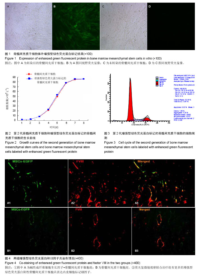
[11] 李丹,朱康顺,周斌,等.慢病毒介导的增强型绿色荧光蛋白报告基因对间充质干细胞的标记[J].中华医学杂志,2010,90(19): 1357-1361.
[12] Herity NA, Lo ST, Oei F,et al.Selective regional myocardial infiltration by the percutaneous coronary venous route: A novel technique for local drug delivery.Catheter Cardiovasc Interv. 2000;51(3):358-363.
[13] Dib N, Menasche P, Bartunek JJ,et al. Recommendations for successful training on methods of delivery of biologics for cardiac regeneration: a report of the International Society for Cardiovascular Translational Research.JACC Cardiovasc Interv. 2010;3(3):265-275.
[14] Wu K, Mo X, Lu S,et al. Retrograde delivery of stem cells: promising delivery strategy for myocardial regenerative therapy. Clin Transplant. 2011;25(6):830-833.
[15] Jain AK, Smith EJ, Rothman MT.The coronary venous system: an alternative route of access to the myocardium.J Invasive Cardiol. 2006;18(11):563-568.
[16] Fearon WF, Ikeno F, Bailey LR,et al. Evaluation of high-pressure retrograde coronary venous delivery of FGF-2 protein.Catheter Cardiovasc Interv. 2004;61(3):422-428.
[17] Yokoyama S, Fukuda N, Li Y,et al. A strategy of retrograde injection of bone marrow mononuclear cells into the myocardium for the treatment of ischemic heart disease.J Mol Cell Cardiol. 2006;40(1):24-34.
[18] Sato T, Iso Y, Uyama T,et al. Coronary vein infusion of multipotent stromal cells from bone marrow preserves cardiac function in swine ischemic cardiomyopathy via enhanced neovascularization.Lab Invest. 2011;91(4):553-564.
[19] Silva GV, Litovsky S, Assad JA,et al. Mesenchymal stem cells differentiate into an endothelial phenotype, enhance vascular density, and improve heart function in a canine chronic ischemia model.Circulation. 2005;111(2):150-156.
[20] Perin EC, Silva GV, Assad JA,et al. Comparison of intracoronary and transendocardial delivery of allogeneic mesenchymal cells in a canine model of acute myocardial infarction.J Mol Cell Cardiol. 2008;44(3):486-495.
[21] Tang J, Xie Q, Pan G,et al. Mesenchymal stem cells participate in angiogenesis and improve heart function in rat model of myocardial ischemia with reperfusion.Eur J Cardiothorac Surg. 2006;30(2):353-361.
[22] Ahn HJ, Lee WJ, Kwack K,et al. FGF2 stimulates the proliferation of human mesenchymal stem cells through the transient activation of JNK signaling.FEBS Lett. 2009; 583 (17): 2922-2926.
[23] Haider HK, Akbar SA, Ashraf M. Angiomyogenesis for myocardial repair.Antioxid Redox Signal. 2009;11(8): 1929-1944.
[24] 杨进福,周文武,唐滔,等.血管内皮生长因子转染骨髓间充质干细胞心肌移植对心肌梗死后大鼠心功能及血管新生的作用[J].中华医学杂志,2006,86(15):1027-1034.
[25] Gao F, He T, Wang H,et al. A promising strategy for the treatment of ischemic heart disease: Mesenchymal stem cell-mediated vascular endothelial growth factor gene transfer in rats.Can J Cardiol. 2007;23(11):891-898. |
.jpg)