中国组织工程研究 ›› 2014, Vol. 18 ›› Issue (28): 4510-4516.doi: 10.3969/j.issn.2095-4344.2014.28.014

• 干细胞培养与分化 stem cell culture and differentiation • 上一篇    下一篇

人牙周膜干细胞与牙周膜细胞生物学特性的比较

封  艳1,粱学萍1,赵  今1,孙玉亮1,钟良军2   

  1. 1新疆医科大学第一附属医院口腔医学中心,新疆维吾尔自治区乌鲁木齐市  830013; 2杭州师范大学附属医院(杭州市第二人民医院)口腔科,浙江省杭州市  310015
  • 出版日期:2014-07-02 发布日期:2014-07-02
  • 通讯作者: 赵今,教授,博士生导师,新疆医科大学第一附属医院口腔医学中心,新疆维吾尔自治区乌鲁木齐市 830013
  • 作者简介:封艳,女, 1973年生,浙江省平湖市人,汉族,新疆医科大学第一附属医院在读博士,主要从事牙周病的治疗研究。
  • 基金资助:

    新疆维吾尔自治区自然科学基金面上项目(2010211A49)

Differences between biological characteristics of human periodontal ligament stem cells and human periodontal ligament cells

Feng Yan1, Liang Xue-ping1, Zhao Jin1, Sun Yu-liang1, Zhong Liang-jun2   

  1. 1 Oral Medicine Center, First Affiliated Hospital of Xinjiang Medical University, Urumqi 830054, Xinjiang Uygur Autonomous Region, China; 2 Affiliated Hospital of Hangzhou Normal University (Second People’s Hospital of Hangzhou), Hangzhou 310015, Zhejiang Province, China
  • Online:2014-07-02 Published:2014-07-02
  • Contact: Zhao Jin, Professor, Doctoral supervisor, Oral Medicine Center, First Affiliated Hospital of Xinjiang Medical University, Urumqi 830054, Xinjiang Uygur Autonomous Region, China
  • About author:Feng Yan, Studying for doctorate, Oral Medicine Center, First Affiliated Hospital of Xinjiang Medical University, Urumqi 830054, Xinjiang Uygur Autonomous Region, China
  • Supported by:

    the Natural Science Foundation of Xinjiang Uygur Autonomous Region, No. 2010211A49

摘要:

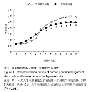
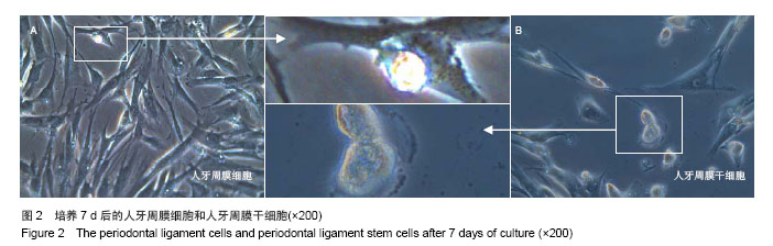
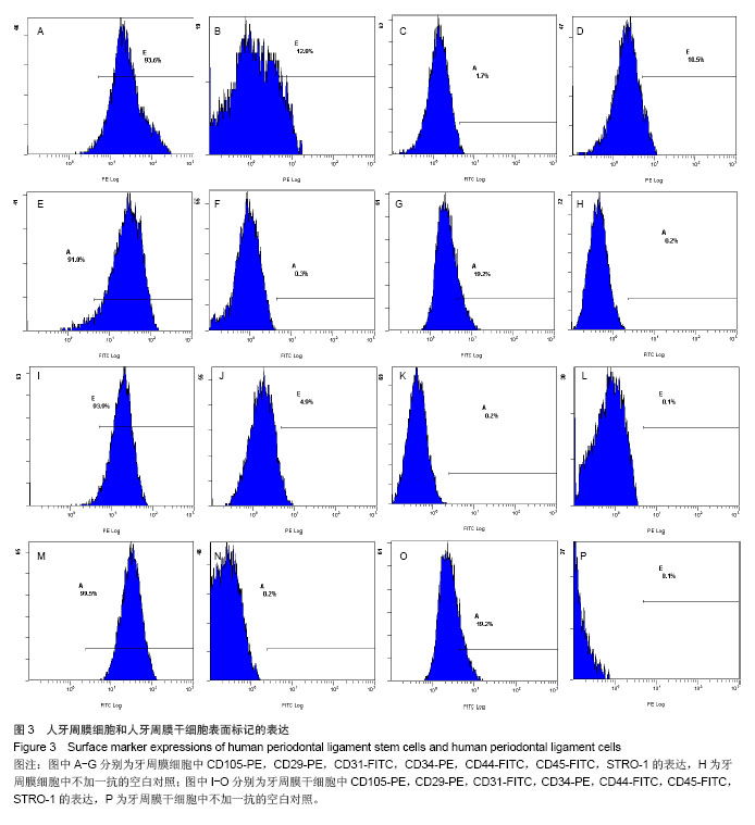
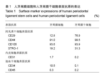
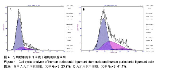
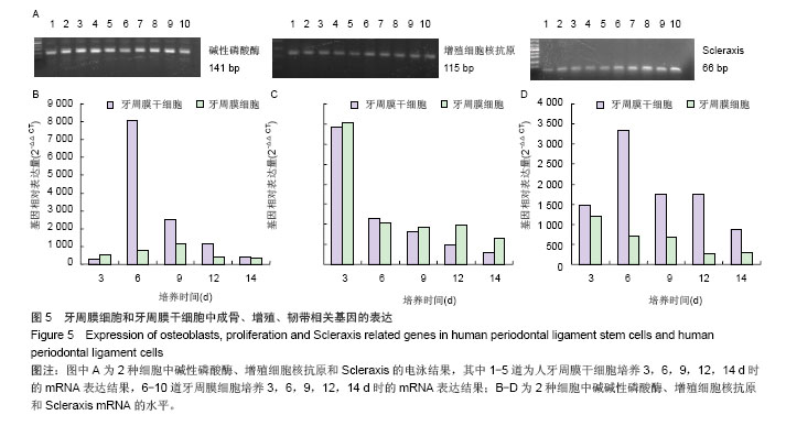
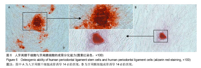
背景:牙周膜干细胞生物作用是目前牙周病治疗研究的热点,牙周膜成纤维细胞是其分化的终末功能细胞之一,也是其主要的支持细胞,两者生物学特性的差异研究鲜有报道。
目的:比较牙周膜干细胞与牙周膜细胞生物学特性的差异。
方法:用组织块法体外对牙周膜细胞以及单细胞克隆分离纯化后的人牙周膜干细胞两种细胞分别进行显微镜下形态观察,CCK8法检测并绘制2种细胞的生长曲线。流式细胞分析比较2种细胞的细胞周期以及细胞表面标记物的表达、实时PCR对2种细胞碱性磷酸酶、增殖细胞核抗原和Scleraxis基因进行检测。
结果与结论:牙周膜干细胞与牙周膜成纤维细胞外观差别明显,人牙周膜干细胞的生长曲线培养前5 d要低于牙周膜细胞,但在5 d后明显高于牙周膜细胞。人牙周膜干细胞与牙周膜细胞的细胞周期分别为41.1%和23.9%。表面标记物检测结果显示2种细胞虽有相似的表达,但在表达率差异有有显著性意义。实时荧光定量PCR结果显示,人牙周膜干细胞在碱性磷酸酶、增殖细胞核抗原以及Scleraxis基因的表达检测均高于牙周膜细胞。表明牙周膜干细胞在成骨增殖等生物学功能上比牙周膜细胞具有更强的潜能。


中国组织工程研究杂志出版内容重点:干细胞;骨髓干细胞;造血干细胞;脂肪干细胞;肿瘤干细胞;胚胎干细胞;脐带脐血干细胞;干细胞诱导;干细胞分化;组织工程


全文链接:

关键词: 干细胞, 培养, 牙周膜干细胞, 牙周膜细胞, 成骨, 增殖, 生物学特性, 表面标志物, 新疆维吾尔自治区自然科学基金

Abstract:

BACKGROUND: The biological function of human periodontal ligament stem cells is a hot area of research in the treatment of periodontal disease. Human periodontal ligament cells are one of the end cells derived from human periodontal ligament stem cells; meanwhile, it can also provide supports to the development of human periodontal ligament stem cells. However, few studies are reported about the difference of biological characteristics between human periodontal ligament stem cells and human periodontal ligament cells.
OBJECTIVE: To compare the differences of biological characteristics between human periodontal ligament stem cells and human periodontal ligament cells.
METHODS: The human periodontal ligament stem cells and human periodontal ligament cells were isolated and purified using tissue explant method and cell clone method, respectively, and then were observed under light microscope to compare the differences of morphology. Cell proliferation curves of human periodontal ligament stem cells and human periodontal ligament cells were drawn respectively with cell counting kit 8 assay. Flow cytometry analysis was used to detect their cell circles and their surface markers expressions. The alkaline phosphatase gene, proliferating cell nuclear antigen gene and Scleraxis gene of human periodontal ligament stem cells and human periodontal ligament cells were detected by Real-time PCR assay. 
RESULTS AND CONCLUSION: The human periodontal ligament stem cells and human periodontal ligament cells showed a notable difference in morphology under the light microscope observation. During the first 5 days, the cell proliferation curve of human periodontal ligament stem cells was lower than that of human periodontal ligament cells, but 5 days later, the curve of human periodontal ligament stem cells was significantly higher than that of human periodontal ligament cells. The cell circles of human periodontal ligament stem cells and human periodontal ligament cells were 41.1% and 23.9%, respectively. The surface markers of human periodontal ligament stem cells and human periodontal ligament cells were similar, but their expression rates had significant difference. The expressions of alkaline phosphatase gene, proliferating cell nuclear antigen gene and Scleraxis gene of human periodontal ligament stem cells were significantly higher than those of human periodontal ligament cells. The above results suggest that human periodontal ligament stem cells have much stronger potential ability than human periodontal ligament cells in osteogensis and cell proliferation.


中国组织工程研究杂志出版内容重点:干细胞;骨髓干细胞;造血干细胞;脂肪干细胞;肿瘤干细胞;胚胎干细胞;脐带脐血干细胞;干细胞诱导;干细胞分化;组织工程


全文链接:

Key words: periodontal ligament, stem cells, cell proliferation, cell differentiation

中图分类号: