| [1]Kadota Y, Yagi H, Inomata K, et al. Mesenchymal stem cells support hepatocyte function in engineered liver grafts. Organogenesis. 2014;10(2).
[2]Nagaishi K, Ataka K, Echizen E, et al. Mesenchymal stem cell therapy ameliorates diabetic hepatocyte damage in mice by inhibiting infiltration of bone marrow-derived cells. Hepatology. 2014;59(5):1816-1829.
[3]Xagorari A, Siotou E, Yiangou M, et al. Protective effect of mesenchymal stem cell-conditioned medium on hepatic cell apoptosis after acute liver injury.Int J Clin Exp Pathol. 2013; 6(5):831-840.
[4]Esrefoglu M. Role of stem cells in repair of liver injury: experimental and clinical benefit of transferred stem cells on liver failure. World J Gastroenterol. 2013;19(40): 6757-6773.
[5]Salomone F, Barbagallo I, Puzzo L, et al. Efficacy of adipose tissue-mesenchymal stem cell transplantation in rats with acetaminophen liver injury. Stem Cell Res. 2013;11(3): 1037-1044.
[6]Wang HS, Hung SC, Peng ST, et al. Mesenchymal stem cells in the Wharton's jelly of the human umbilical cord. Stem Cells. 2004;22(7):1330-1337.
[7]Kumar M, Sarin SK. Is cirrhosis of the liver reversible? Indian J Pediatr. 2007;74(4):393-399.
[8]Hong J, Jin H, Han J, et al. Infusion of human umbilical cord derived mesenchymal stem cells effectively relieves liver cirrhosis in DEN induced rats. Mol Med Rep. 2014;9(4): 1103-1111.
[9]Poynard T, McHutchison J, Manns M, et al. Impact of pegylated interferon alfa-2b and ribavirin on liver fibrosis in patients with chronic hepatitis C. Gastroenterology. 2002; 122(5):1303-1313.
[10]Shepherd J, Brodin H, Cave C,et al.Pegylated interferon alpha-2a and -2b in combination with ribavirin in the treatment of chronic hepatitis C: a systematic review and economic evaluation.Health Technol Assess. 2004;8(39):iii-iv, 1-125.
[11]Toshikuni N, Arisawa T, Tsutsumi M.Hepatitis C-related liver cirrhosis - strategies for the prevention of hepatic decompensation, hepatocarcinogenesis, and mortality.World J Gastroenterol. 2014;20(11):2876-2887.
[12]Terai S, Takami T, Yamamoto N, et al. Status and Prospects of Liver Cirrhosis Treatment by Using Bone Marrow-Derived Cells and Mesenchymal Cells. Tissue Eng Part B Rev. 2014 Mar 7.
[13]Wanless IR. Use of corticosteroid therapy in autoimmune hepatitis resulting in the resolution of cirrhosis. J Clin Gastroenterol. 2001;32(5):371-372.
[14]Shao CH, Chen SL, Dong TF, et al. Transplantation of bone marrow-derived mesenchymal stem cells after regional hepatic irradiation ameliorates thioacetamide-induced liver fibrosis in rats.J Surg Res. 2014;186(1):408-416.
[15]Li T, Zhu J, Ma K,et al. Autologous bone marrow-derived mesenchymal stem cell transplantation promotes liver regeneration after portal vein embolization in cirrhotic rats. J Surg Res. 2013;184(2):1161-1173.
[16]Motawi TM, Atta HM, Sadik NA, et al. The therapeutic effects of bone marrow-derived mesenchymal stem cells and simvastatin in a rat model of liver fibrosis. Cell Biochem Biophys. 2014;68(1):111-125.
[17]Fang B, Shi M, Liao L, et al. Systemic infusion of FLK1(+) mesenchymal stem cells ameliorate carbon tetrachloride-induced liver fibrosis in mice. Transplantation. 2004;78(1):83-88.
[18]Lee KD, Kuo TK, Whang-Peng J, et al. In vitro hepatic differentiation of human mesenchymal stem cells. Hepatology. 2004;40(6):1275-1284.
[19]Sakaida I,Terai S,Nishina H,et al.Development of cell therapy using autologous bone marrow cells for liver cirrhosis. Med Mol Morphol. 2005;38(4):197-202.
[20]Hung SC, Chen NJ, Hsieh SL, et al. Isolation and characterization of size-sieved stem cells from human bone marrow. Stem Cells. 2002;20(3):249-258.
[21]Oe H, Kaido T, Mori A, et al. Hepatocyte growth factor as well as vascular endothelial growth factor gene induction effectively promotes liver regeneration after hepatectomy in Solt-Farber rats. Hepatogastroenterology. 2005;52(65): 1393-1397.
[22]Shi MN, Huang YH, Zheng WD, et al. Relationship between transforming growth factor beta1 and anti-fibrotic effect of interleukin-10. World J Gastroenterol. 2006;12(15): 2357-2362.
[23]Shi MN, Zheng WD, Zhang LJ, et al. Effect of IL-10 on the expression of HSC growth factors in hepatic fibrosis rat. World J Gastroenterol. 2005;11(31):4788-4793.
[24]Zheng WD, Zhang LJ, Shi MN, et al. Expression of matrix metalloproteinase-2 and tissue inhibitor of metalloproteinase-1 in hepatic stellate cells during rat hepatic fibrosis and its intervention by IL-10. World J Gastroenterol. 2005;11(12):1753-1758.
[25]Nan YM, Kong LB, Ren WG, et al.Activation of peroxisome proliferator activated receptor alpha ameliorates ethanol mediated liver fibrosis in mice.Lipids Health Dis. 2013;12:11.
[26]Suominen JS, Lampela H, Heikkilä P,Myofibroblastic cell activation and neovascularization predict native liver survival and development of esophageal varices in biliary atresia. World J Gastroenterol. 2014;20(12):3312-3319.
[27]Perepelyuk M, Terajima M, Wang AY, et al. Hepatic stellate cells and portal fibroblasts are the major cellular sources of collagens and lysyl oxidases in normal liver and early after injury. Am J Physiol Gastrointest Liver Physiol. 2013;304(6): G605-614. |
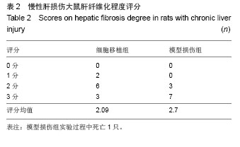
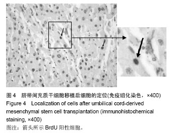
.jpg)