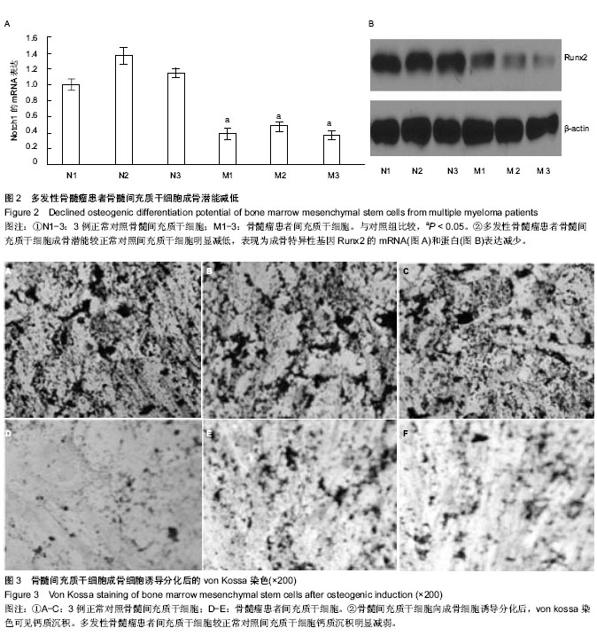
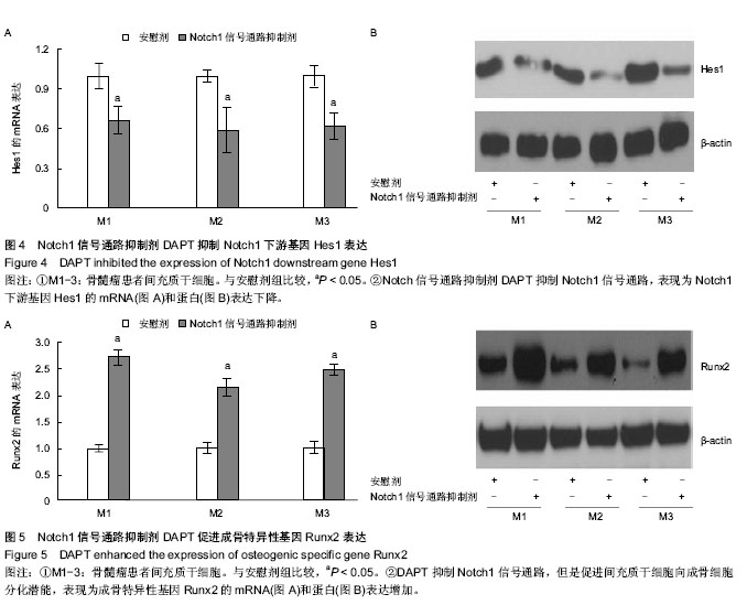
| [1] Alegre A, Gironella M, Bailén A, et al. Zoledronic acid in the management of bone disease as a consequence of multiple myeloma: a review. Eur J Haematol. 2013 Dec 14.
[2] Giuliani N, Bataille R, Mancini C, et al. Myeloma cells induce imbalance in the osteoprotegerin/osteoprotegerin ligand system in the human bone marrow environment. Blood. 2001; 98(13):3527-3533.
[3] Giuliani N, Morandi F, Tagliaferri S, et al. The proteasome inhibitor bortezomib affects osteoblast differentiation in vitro and in vivo in multiple myeloma patients. Blood. 2007;110(1): 334-338.
[4] Horwitz EM, Le Blanc K, Dominici M, et al. Clarification of the nomenclature for MSC: The International Society for Cellular Therapy position statement. Cytotherapy. 2005;7(5):393-395.
[5] Reagan MR, Ghobrial IM. Multiple myeloma mesenchymal stem cells: characterization, origin, and tumor-promoting effects. Clin Cancer Res. 2012;18(2):342-349.
[6] Giuliani N, Mangoni M, Rizzoli V. Osteogenic differentiation of mesenchymal stem cells in multiple myeloma: identification of potential therapeutic targets. Exp Hematol. 2009;37(8):879-886.
[7] Wharton KA, Yedvobnick B, Finnerty VG, et al. opa: a novel family of transcribed repeats shared by the Notch locus and other developmentally regulated loci in D. melanogaster. Cell. 1985;40(1):55-62.
[8] Xu N, Liu H, Qu F, et al. Hypoxia inhibits the differentiation of mesenchymal stem cells into osteoblasts by activation of Notch signaling. Exp Mol Pathol. 2013;94(1):33-39.
[9] Criteria for the classification of monoclonal gammopathies, multiple myeloma and related disorders: a report of the International Myeloma Working Group. Br J Haematol. 2003; 121(5):749-757.
[10] Chen Z, Orlowski RZ, Wang M, et al. Osteoblastic niche supports the growth of quiescent multiple myeloma cells. Blood. 2014;123(14):2204-2208.
[11] Sezer O. Myeloma bone disease: recent advances in biology, diagnosis, and treatment. Oncologist. 2009;14(3):276-283.
[12] Galson DL, Silbermann R, Roodman GD. Mechanisms of multiple myeloma bone disease. Bonekey Rep. 2012;1:135.
[13] Roodman GD. Osteoblast function in myeloma. Bone. 2011; 48(1):135-140.
[14] Giuliani N, Rizzoli V. Myeloma cells and bone marrow osteoblast interactions: role in the development of osteolytic lesions in multiple myeloma. Leuk Lymphoma. 2007;48(12): 2323-2329.
[15] Zhou F, Meng S, Song H, et al. Dickkopf-1 is a key regulator of myeloma bone disease: opportunities and challenges for therapeutic intervention. Blood Rev. 2013;27(6):261-267.
[16] Kristensen IB, Christensen JH, Lyng MB, et al. Expression of osteoblast and osteoclast regulatory genes in the bone marrow microenvironment in multiple myeloma: only up-regulation of Wnt inhibitors SFRP3 and DKK1 is associated with lytic bone disease. Leuk Lymphoma. 2014; 55(4):911-919.
[17] Tsirakis G, Roussou P, Pappa CA, et al. Increased serum levels of MIP-1alpha correlate with bone disease and angiogenic cytokines in patients with multiple myeloma. Med Oncol. 2014;31(1):778.
[18] Giuliani N, Colla S, Morandi F, et al. Myeloma cells block RUNX2/CBFA1 activity in human bone marrow osteoblast progenitors and inhibit osteoblast formation and differentiation. Blood. 2005;106(7):2472-2483.
[19] Mohty M, Malard F, Mohty B, et al. The effects of bortezomib on bone disease in patients with multiple myeloma. Cancer. 2014;120(5):618-623.
[20] Qiang YW, Heuck CJ, Shaughnessy JD, et al. Proteasome inhibitors and bone disease. Semin Hematol. 2012;49(3): 243-248.
[21] Zangari M, Terpos E, Zhan F, et al. Impact of bortezomib on bone health in myeloma: a review of current evidence. Cancer Treat Rev. 2012;38(8):968-980.
[22] Mukherjee S, Raje N, Schoonmaker JA, et al. Pharmacologic targeting of a stem/progenitor population in vivo is associated with enhanced bone regeneration in mice. J Clin Invest. 2008; 118(2):491-504.
[23] Qiang YW, Hu B, Chen Y, et al. Bortezomib induces osteoblast differentiation via Wnt-independent activation of beta-catenin/TCF signaling. Blood. 2009;113(18):4319-4330.
[24] Tan T, Lu B, Zhang J, et al. Notch1 Signaling Antagonizes Transforming Growth Factor-β Pathway and Induces Apoptosis in Rabbit Trophoblast Stem Cells. Stem Cells Dev. 2014;23(8):813-822.
[25] Mirandola L, Apicella L, Colombo M, et al. Anti-Notch treatment prevents multiple myeloma cells localization to the bone marrow via the chemokine system CXCR4/SDF-1. Leukemia. 2013;27(7):1558-1566.
[26] Guo D, Li C, Teng Q, et al. Notch1 overexpression promotes cell growth and tumor angiogenesis in myeloma. Neoplasma. 2013;60(1):33-40.
[27] Nefedova Y, Cheng P, Alsina M, et al. Involvement of Notch-1 signaling in bone marrow stroma-mediated de novo drug resistance of myeloma and other malignant lymphoid cell lines. Blood. 2004;103(9):3503-3510.
[28] Boopathy AV, Pendergrass KD, Che PL, et al. Oxidative stress-induced Notch1 signaling promotes cardiogenic gene expression in mesenchymal stem cells. Stem Cell Res Ther. 2013;4(2):43.
[29] Hilton MJ, Tu X, Wu X, et al. Notch signaling maintains bone marrow mesenchymal progenitors by suppressing osteoblast differentiation. Nat Med. 2008;14(3):306-314.
[30] Jung SR, Song NJ, Yang DK, et al. Silk proteins stimulate osteoblast differentiation by suppressing the Notch signaling pathway in mesenchymal stem cells. Nutr Res. 2013; 33(2): 162-170.
[31] Schwarzer R, Kaiser M, Acikgoez O, et al. Notch inhibition blocks multiple myeloma cell-induced osteoclast activation. Leukemia. 2008;22(12):2273-2277. |