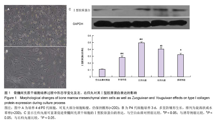
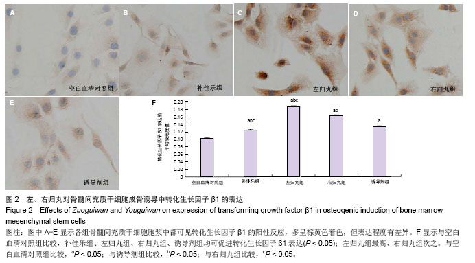
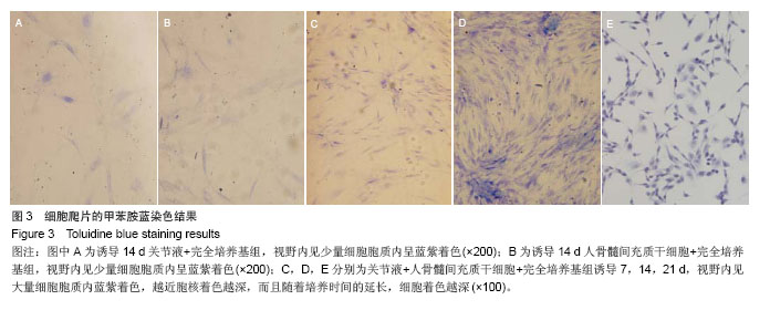
| [1]Pittenger MF, Mackay AM, Beck SC, et al. Multilineage potential of adult human mesenchymal stem cells. Science. 1999;284(5411):143-147.[2]Zheng YH, Xiong W, Su K, et al. Multilineage differentiation of human bone marrow mesenchymal stem cells in vitro and in vivo. Exp Ther Med. 2013;5(6):1576-1580.[3]张君红,姜海行,覃山羽,等.全骨髓细胞贴壁法分离与培养大鼠骨髓间充质干细胞的多向分化能力[J].中国组织工程研究,2012, 16(36):6685-6689.[4]Xu S, Xu YC. Recent progress of BMSCs acting as seeding cell for tissue engineered cartilage. Zhongguo Xiu Fu Chong Jian Wai Ke Za Zhi. 2008;22(2):163-166.[5]王丽雅,廖学娟,郑晓辉,等.Wnt信号通路与骨髓间充质干细胞成骨之间的关系[J].现代生物医学进展,2012,20(12):3981-3984.[6]Chen ZX, Wang XF, Shao YC, et al. Synthetic osteogenic growth peptide promotes differentiation of human bone marrow mesenchymal stem cells to osteoblasts via RhoA/ROCK pathway. Mol Cell Biochem. 2011;358(1-2):221-227.[7]Zheng YH, Xiong W, Su K, et al. Multilineage differentiation of human bone marrow mesenchymal stem cells in vitro and in vivo. Exp Ther Med.2013;5(6):1576-1580.[8]Lee J, Vasikaran S. Current recommendations for laboratory testing and use of bone turnover markers in management of osteoporosis. Ann Lab Med. 2012;32(2):105-112.[9]吕沐瀚,李晓云,李吕平等.转化生长因子β1对肝星状细胞活化及跨膜信号转导影响与疏肝颗粒的干预[J].中国组织工程研究与临床康复,2009,13(50):9898-9902.[10]蔡建平,张爱国,许宝满.五种中药(或方剂)含药血清对体外骨髓间充质干细胞增殖活性影响的筛选研究[J].时珍国医国药,2011, 22(6):1385-1387.[11]Liu M, Xiao GG, Ron P, et al. Semen Astragali Complanati- and Rhizoma Cibotii-enhanced bone formation in osteoporosis rat. BMC Complement Altern Med.2013; 20(13):141.[12]杨锋,唐德志,卞琴,等.中药诱导骨髓间充质干细胞的成骨分化[J].中国组织工程研究与临床康复,2011,15(10):1847-1850.[13]Chen WH, Wang HM. Experimental research progress of warming yang and reinforcing kidney of Chinese medicine to promote the differentiation of bone marrow stromal cells. Zhongguo Gu Shang. 2011;24(4):352-356.[14]Bian Q, Huang JH, Liu SF, et al. Different molecular targets of Icariin on bMSCs in CORT and OVX –rats. Front Biosci (Elite Ed). 2012;4(1):1224-1236.[15]宋囡,何文智,王智民,等.左、右归丸及其拆方对骨髓间充质干细胞成骨分化的影响[J].中国病理生理杂志,2013,29(7):1268-1274.[16]周岩,朴金花,金莲花,等.骨髓间充质干细胞在心血管疾病中的研究及应用[J].中国组织工程研究,2013,17(1):112-117.[17]Panetta NJ, Gupta DM, Longaker MT. Bone regeneration and repair. Curr Stem Cell Res Ther. 2010;5(2):122-128.[18]Derubeis AR, Cancedda R. Bone marrow stromal cells (BMSCs) in bone engineering: limitations and recent advances.Ann Biomed Eng. 2004;32(1):160-165.[19]蒿长英,任艳玲,刘立萍,等.左归丸含药血清通过ERK/TGF-β/ Smads信号级联调控MC3T3-E1细胞增殖与分化[J].中国病理生理杂志,2012,28(9):1670-1675.[20]王艳杰,任艳玲,宋囡,等.左归丸对去卵巢大鼠股骨中Ⅰ型胶原mRNA表达的影响[J].中国老年学杂志,2012,32(12):5170-5172.[21]Liu LP, Ran L, Ren YL, et al. Zuogui Pill stimulates Runx2 expression in MC3T3 cells by p38 MAPK signaling pathways. CJTCMP.2013;28(5):1457-1461.[22]Gelse K,Poschl E,Aigner T. Collagen-structure, function, and biosynthesis. Adv Drug Delivery Rec. 2003;55:1531-1546.[23]Oliveira MR, Martins Ed, Célio-Mariano R, et al. Tissue engineering: using collagen type I matrix for bone healing of bone defects. J Craniofacial Surg. 2013;24(4):394-396.[24]原超,徐无忌,陈映娟.大鼠骨髓间充质干细胞与成骨细胞共育环境下Ⅰ型胶原的表达[J].中国组织工程研究与临床康复,2009, 13(33):6423-6427.[25]汤小康,应航,李敏,等.Ⅰ型与Ⅱ型胶原酶在原代成骨细胞培养中消化效果的对比研究[J].中医正骨,2013,25(6):406-409.[26]余希杰,杨志明,屈艺,等.Ⅰ型胶原及其受体系统在成骨细胞内的表达[J].中国修复重建外科杂志,2000,14(4):234-236.[27]Zi Z, Chapnick DA, Liu X. Dynamics of TGF-β/Smad signaling. FEBS Letters. 2012;586(7):1921-1928.[28]Massagué J. TGFβ signalling in context. Nat Rev Mol Cell Biol. 2012;13(10):616-630.[29]Zou QP, Yang E, Zhang HS. Effect of the methylation enzyme inhibitors of 5-aza-2-deoxycytidine on the TGF-beta/smad signal transduction pathway in human keloid fibroblasts. Zhonghua Zheng Xing Wai Ke Za Zhi. 2013;29(4):285-289.[30]Longmei Zhao, Shu Jiang, Basil M. Hantash. Transforming Growth Factor β1 Induces Osteogenic Differentiation of Murine Bone Marrow Stromal Cells .Tissue Eng Part A. 2010; 16(2):725-733.[31]Hough C, Radu M, DoréJJ. Tgf-beta induced Erk phosphorylation of smad linker region regulates smad signaling. PLoS One. 2012;7(8):e42513.[32]Source Nakao A, Imamura T, Souchelnytskyi S, et al. TGF-beta receptor-mediated signalling through Smad2, Smad3 and Smad4. EMBO J. 1997;16(17):5353-5362.[33]Kawabata M, Miyazono K. Signal transduction of the TGF-beta superfamily by Smad proteins. J Biochem. 1999;125(1):9-16.[34]Massagué J, Seoane J, Wotton D. Smad transcription factors. Genes Dev. 2005;19(23):2783-2810.[35]Xu XL, Dai KR, Tang TT. The role of Smads and related transcription factors in the signal transduction of bone morphogenetic protein inducing bone formation. Chinese Journal of Reparative and Reconstructive Surgery. 2003; 17(5):359-362.[36]Matsuzaki K. Smad phospho-isoforms direct context-dependent TGF-β signaling. Cytokine Growth Factor Rev. 2013;24(4):385- 399.[37]Brown KA,Pietenpol JA,Moses HL.A tale of two proteins: differential roles and regulation of Smad2 and Smad3 in TGF-bate signaling. J Cell Biochem. 2007;101(1):9-33.[38]Makkar P, Metpally RP, Sangadala S, et al. Modeling and analysis of MH1 domain of Smads and their interaction with promoter DNA sequence motif. J Mol Graph Model.2009; 27(7):803-812.[39]Jin Q, Gao G, Mulder KM. A dynein motor attachment complex regulates TGFß/Smad3 signaling. Int J Biol Sci. 2013;9(6):531-540.[40]Ten Dijke P, Hill CS. New insights into TGF-beta-Smad signalling. Trends Biochem Sci.2004;29(5):265-273.[41]Brown KA, Pietenpol JA, Moses HL. A tale of two proteins: differential roles and regulation of Smad2 and Smad3 in TGF-beta signaling. J Cell Biochem. 2007;101(1):9-33.[42]Inman GJ, Nicolás FJ, Hill CS. Nucleocytoplasmic shuttling of Smads2, 3, and 4 permits sensing of TGF-beta receptor activity. Molecular Cell. 2002;10(2):283-294. |
.jpg)
.jpg)