中国组织工程研究 ›› 2014, Vol. 18 ›› Issue (9): 1313-1318.doi: 10.3969/j.issn.2095-4344.2014.09.001

• 骨与关节图像与影像 bone and joint imaging •    下一篇

兔腰椎间盘髓核穿刺抽吸后的影像及组织病理学变化

刘航涛1,王万明2,林智军1,陈国仙3,林国叶1,李平生1   

  1. 1解放军南京军区福州总院第一附属医院骨科,福建省莆田市  351100;2解放军南京军区福州总医院骨科,福建省福州市  350025;3莆田市第一医院骨科,福建省莆田市  351100
  • 出版日期:2014-02-26 发布日期:2014-02-26
  • 通讯作者: 王万明,博士,主任医师,硕士生导师,解放军南京军区福州总医院骨科,福建省福州市 350025
  • 作者简介:刘航涛,男,1982年生,河南省周口市人,汉族,2008年福建中医学院毕业,硕士,主治医师,主要从事创伤临床及脊柱外科研究工作。
  • 基金资助:

    莆田市科技计划项目(2010 S09-2)

Alterations in imaging and histopathology after aspiration of nucleus pulposus of rabbit lumbar intervertebral disc

Liu Hang-tao1, Wang Wan-ming2, Lin Zhi-jun1, Chen Guo-xian3, Lin Guo-ye1, Li Ping-sheng1   

  1. 1 Department of Orthopedics, the First Hospital Affiliated to Fuzhou General Hospital of Nanjing Military Area Command of Chinese PLA, Putian 351100, Fujian Province, China; 2 Department of Orthopedics, Fuzhou General Hospital of Nanjing Military Area Command of Chinese PLA, Fuzhou 350025, Fujian Province, China; 3 Department of Orthopedics, Putian Municipal First Hospital, Putian 351100, Fujian Province, China
  • Online:2014-02-26 Published:2014-02-26
  • Contact: Wang Wan-ming, M.D., Chief physician, Master’s supervisor, Department of Orthopedics, Fuzhou General Hospital of Nanjing Military Area Command of Chinese PLA, Fuzhou 350025, Fujian Province, China
  • About author:Liu Hang-tao, Master, Attending physician, Department of Orthopedics, the First Hospital Affiliated to Fuzhou General Hospital of Nanjing Military Area Command of Chinese PLA, Putian 351100, Fujian Province, China
  • Supported by:

    the Putian Municipal Science and Technology Project, No.2010 S09-2

摘要:

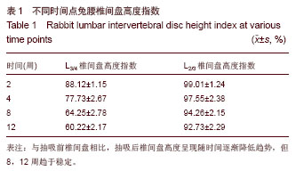
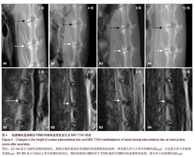
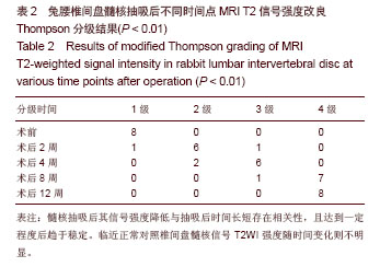
背景:髓核摘除后椎间盘会随时间出现什么样的影像学及组织病理学变化,目前尚不明确。
目的:观察兔腰椎间盘髓核穿刺抽吸术后影像学及组织病理学的变化。
方法:32只日本大耳白兔,用21号针头行L 3/4椎间盘后外侧穿刺抽吸出部分髓核组织,L 2/3椎间盘作为正常对照椎间盘,于抽吸后2,4,8,12周时按照分组取8只兔子行腰椎侧位X射线检查,测量L 3/4 、L 2/3椎间隙高度并计算椎间盘高度指数,行正中矢状位MRI检查及椎间盘组织病理学检查。
结果与结论:髓核抽吸后2,4,8,12周椎间盘高度呈逐渐降低趋势,但8-12周变化减小,与正常对照组椎间盘相比,各时间点椎间盘高度指数显著降低(P < 0.05)。抽吸后2,4,8,12周的髓核信号强度随时间逐渐降低,8周时已达改良Thompson分级标准的4级。抽吸后凝胶状髓核组织随时间逐渐出现裂隙,形态逐渐紊乱,12周时呈现明显的纤维化表现,髓核4周时出现较多的类软骨细胞,呈现活跃状态,髓核细胞明显减少,抽吸后8,12周髓核内纤维样细胞增多,类软骨细胞数量减少,纤维环随时间逐渐出现扭曲,排列紊乱,突起,出现分层、纤维断裂现象。说明后外侧纤维环穿刺髓核抽吸后,兔腰椎间盘X射线高度、MRI T2加权信号强度随时间逐渐降低、减弱,椎间盘组织逐渐出现退变病理改变,但8-12周其变化趋于缓和。


中国组织工程研究杂志出版内容重点:人工关节;骨植入物;脊柱骨折;内固定;数字化骨科;组织工程


全文链接:

关键词: 植入物, 脊柱植入物, 椎间盘, 退变, 髓核, 影像学, 椎间盘高度, 椎间盘高度指数, 病理学

Abstract:

BACKGROUND: It remains unclear what kind of changes in imaging and histopathology would appear in intervertebral disc after removal of nucleus pulposus over time.
OBJECTIVE: To observe the alteration in imaging and histopathology of rabbit lumbar intervertebral disc after nucleus pulposus aspiration.
METHODS: A total of 32 Japanese rabbits were subjected to nucleus pulposus aspiration in posterior lateral L 3/4 intervertebral disc with a 21-gauge hypodermic needle. L 2/3 intervertebral disc served as a normal control. At 2, 4, 8 and 12 weeks after aspiration, eight rabbits according to grouping received radiograph in the lateral position of lumbar vertebra. The height of L 3/4 and L 2/3 intervertebral space was measured and intervertebral disc height index was calculated. Rabbits received median sagittal MRI examination and histopathological examination of intervertebral disc.
RESULTS AND CONCLUSION: The intervertebral disc height degraded slowly at 2, 4, 8, and 12 weeks after aspiration. However, the difference between 8 and 12 weeks was reduced. Compared with normal control, intervertebral disc height index significantly reduced at various time points (P < 0.05). The nucleus pulposus signal intensity was gradually reduced at 2, 4, 8, and 12 weeks after aspiration, and reached grade 4 of modified Thompson grading criteria at 8 weeks. After aspiration, gelatinous nucleus pulposus of disc slowly exhibited fissures with time, morphous was slowly disordered and showed significant fibrosis performance at 12 weeks. In nucleus pulposus region, more cartilage-like cells were found at 4 weeks after operation. The cells were active. Nucleus pulposus cells decreased significantly. At 8 and 12 weeks after operation, fibroblasts increased in nucleus pulposus region, and the number of cartilage-like cells reduced. The annulus fibrosus gradually became distorted, disordered, with processes and layers, and fiber breakage appeared. These results indicated that after puncturing lateral annulus fibrosus and aspirating the nucleus pulposus, radiograph height of the intervertebral disc and MRI T2-weighted signal intensity gradually reduced. Pathological changes were observed. However, the degeneration alleviated between 8-12 weeks.


中国组织工程研究杂志出版内容重点:人工关节;骨植入物;脊柱骨折;内固定;数字化骨科;组织工程


全文链接:

Key words: intervertebral disk, disk degeneration, radiographic image enhancement, pathology

中图分类号: