| [1] Djouad F, Plence P, Bony C,et al. Immunosuppressive effect of mesenchymal stem cells favors tumor growth in allogeneic animals. Blood. 2003;102(10):3837-3844.
[2] Nauta AJ, Fibbe WE. Immunomodulatory properties of mesenchymal stromal cells. Blood. 2007;110(10):3499-3506.
[3] Ren G, Zhang L, Zhao X,et al. Mesenchymal stem cell-mediated immunosuppression occurs via concerted action of chemokines and nitric oxide. Cell Stem Cell. 2008; 2(2):141-150.
[4] Pittenger MF, Mackay AM, Beck SC,et al. Multilineage potential of adult human mesenchymal stem cells. Science. 1999;284(5411):143-147.
[5] Deng J, Petersen BE, Steindler DA,et al. Mesenchymal stem cells spontaneously express neural proteins in culture and are neurogenic after transplantation.Stem Cells. 2006;24(4): 1054-1064.
[6] Sakuragawa N, Kakinuma K, Kikuchi A,et al. Human amnion mesenchyme cells express phenotypes of neuroglial progenitor cells. J Neurosci Res. 2004;78(2):208-214.
[7] Whittle WL, Gibb W, Challis JR.The characterization of human amnion epithelial and mesenchymal cells: the cellular expression, activity and glucocorticoid regulation of prostaglandin output. Placenta. 2000;21(4):394-401.
[8] In 't Anker PS, Scherjon SA, Kleijburg-van der Keur C,et al. Isolation of mesenchymal stem cells of fetal or maternal origin from human placenta. Stem Cells. 2004;22(7):1338-1345.
[9] Alviano F, Fossati V, Marchionni C,et al. Term Amniotic membrane is a high throughput source for multipotent Mesenchymal Stem Cells with the ability to differentiate into endothelial cells in vitro. BMC Dev Biol. 2007;7:11.
[10] Miki T, Lehmann T, Cai H,et al. Stem cell characteristics of amniotic epithelial cells. Stem Cells. 2005;23(10):1549-1559.
[11] Takashima S, Ise H, Zhao P,et al. Human amniotic epithelial cells possess hepatocyte-like characteristics and functions. Cell Struct Funct. 2004;29(3):73-84.
[12] Miki T, Lehmann T, Cai H,et al. Stem cell characteristics of amniotic epithelial cells. Stem Cells. 2005;23(10):1549-1559.
[13] Luo H, Huang X, Huang F,et al. Transversal inducing differentiation of human amniotic epithelial cells into hepatocyte-like cells. Zhong Nan Da Xue Xue Bao Yi Xue Ban. 2011;36(6):525-531.
[14] Yen BL, Huang HI, Chien CC,et al. Isolation of multipotent cells from human term placenta.Stem Cells. 2005;23(1):3-9.
[15] 方宁,章涛,陈代雄.人羊膜细胞表型可塑性研究进展[J].中国生物工程杂志,2008,28(6s):191-195.
[16] 张路,方宁,陈代雄,等.人羊膜上皮细胞有向心肌样细胞分化的特性[J].中国组织工程研究与临床康复,2008,12(3):401-405.
[17] Pratama G, Vaghjiani V, Tee JY, et al. Changes in culture expanded human amniotic epithelial cells: implications for potential therapeutic applications.PLoS One. 2011;6(11): e26136.
[18] Steed DL, Trumpower C, Duffy D,et al. Amnion-derived cellular cytokine solution: a physiological combination of cytokines for wound healing. Eplasty. 2008;8:e18.
[19] Koh JW, Shin YJ, Oh JY,et al.The expression of TIMPs in cryo-preserved and freeze-dried amniotic membrane.Curr Eye Res. 2007;32(7-8):611-616.
[20] 李娟,王克华,张丽红,等.人类早期胚胎与体细胞共培养的研究进展[J].国际生殖健康/计划生育杂志,2009,28(2):99-102.
[21] Mikos AG, Sarakinos G, Leite SM,et al. Laminated three-dimensional biodegradable foams for use in tissue engineering. Biomaterials. 1993;14(5):323-330.
[22] Ishaug-Riley SL, Crane-Kruger GM, Yaszemski MJ,et al.Three-dimensional culture of rat calvarial osteoblasts in porous biodegradable polymers. Biomaterials. 1998;19(15): 1405-1412.
[23] Holy CE, Yakubovich R. Processing cell-seeded polyester scaffolds for histology. J Biomed Mater Res. 2000;50(2): 276-279.
[24] Langer R, Vacanti JP.Tissue engineering. Science. 1993; 260(5110):920-926. |
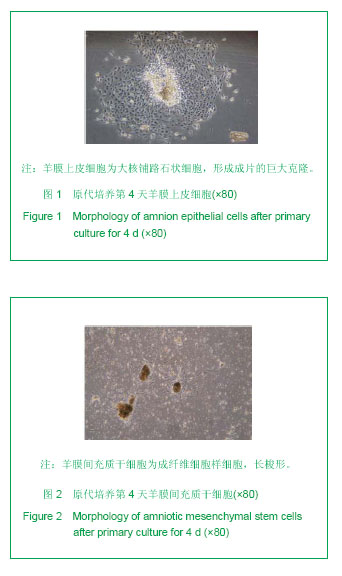
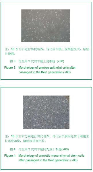
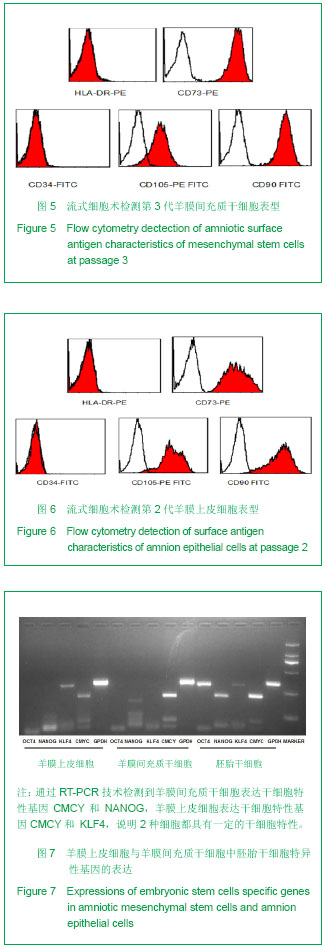
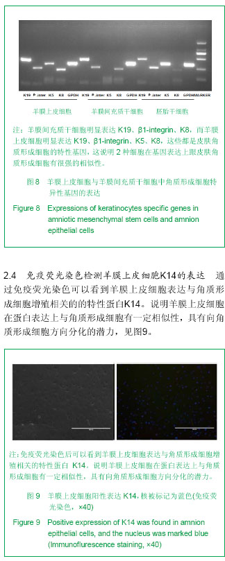
.jpg)
.jpg)