中国组织工程研究 ›› 2020, Vol. 24 ›› Issue (22): 3460-3466.doi: 10.3969/j.issn.2095-4344.2299

• 药物控释材料 drug delivery materials • 上一篇    下一篇

水凝胶三维培养对人羊膜间充质干细胞特性及旁分泌效应的影响

王  旗,杨晓双,王达利   

  1. 遵义医科大学附属医院整形外科,贵州省遵义市  563000
  • 收稿日期:2019-11-11 修回日期:2019-12-02 接受日期:2020-01-08 出版日期:2020-08-08 发布日期:2020-04-26
  • 通讯作者: 王达利,教授,硕士生导师,遵义医科大学附属医院整形外科,贵州省遵义市 563000
  • 作者简介:王旗,女,1993年生,山东省济宁市人,汉族,遵义医科大学在读硕士,主要从事干细胞与创面愈合相关研究。 共同第一作者:杨晓双,女,1992年生,新疆维吾尔自治区石河子市人,汉族,医师,主要从事创面修复与整形研究。
  • 基金资助:
    国家自然科学基金项目(81560313);国家自然科学基金面上项目(81871570)

Effects of hydrogel three-dimensional culture on the characteristics of human amniotic mesenchymal stem cells and paracrine effect

Wang Qi, Yang Xiaoshuang, Wang Dali   

  1. Department of Plastic Surgery, Affiliated Hospital of Zunyi Medical University, Zunyi 563000, Guizhou Province, China
  • Received:2019-11-11 Revised:2019-12-02 Accepted:2020-01-08 Online:2020-08-08 Published:2020-04-26
  • Contact: Wang Dali, Professor, Master’ supervisor, Department of Plastic Surgery, Affiliated Hospital of Zunyi Medical University, Zunyi 563000, Guizhou Province, China
  • About author:Wang Qi, Master candidate, Department of Plastic Surgery, Affiliated Hospital of Zunyi Medical University, Zunyi 563000, Guizhou Province, China Yang Xiaoshuang, Physician, Department of Plastic Surgery, Affiliated Hospital of Zunyi Medical University, Zunyi 563000, Guizhou Province, China Wang Qi and Yang Xiaoshuang contributed equally to the study.
  • Supported by:
    the National Natural Science Foundation of China, No. 81560313; the National Natural Science Foundation of China (General Program), No. 81871570

摘要:

文题释义:

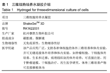
水凝胶三维培养:应用安全稳定的天然或合成聚合物材料为细胞提供一个空间、立体的生存环境,可增强细胞的黏附、增殖及分泌细胞因子能力。

旁分泌效应:以往研究认为干细胞应用于创面治疗的潜能是干细胞归巢分化为损伤组织,后来发现移植的干细胞大多停留在肝、脾,很少能到达损伤创面。目前研究证实间充质干细胞通过分泌某些营养因子作用于临近的靶细胞,起到促进创面愈合的作用。

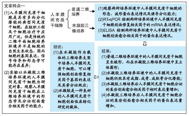
背景:研究表明间充质干细胞在创面愈合中具有减轻炎症反应、促进创面愈合、减轻瘢痕形成的作用,然而以往的普通二维培养环境由于存在细胞间接触性抑制,可能导致细胞的基因表达、信号传导和形态学存在差异。

目的:观察人羊膜间充质干细胞旁分泌创面愈合相关因子的能力是否受二维培养环境与三维培养环境的影响。

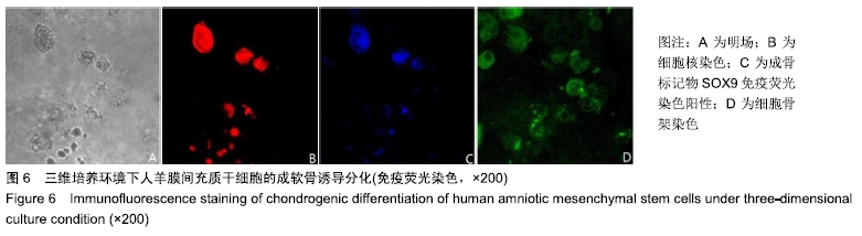
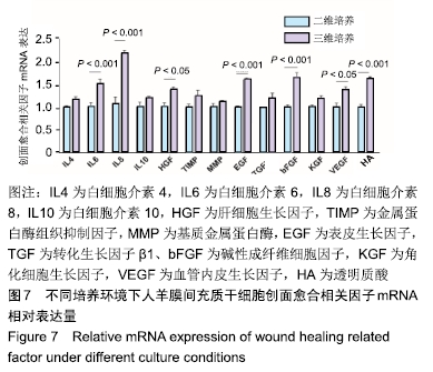
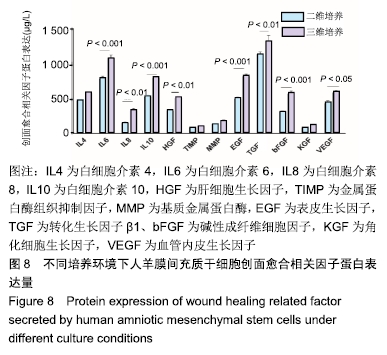
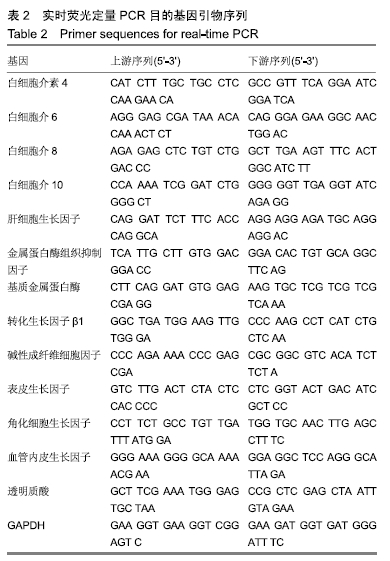
方法:利用传统酶消化法获得人羊膜间充质干细胞后,分别接种于普通细胞培养瓶(二维培养)与ShakeGelTM 3D水凝胶中(三维培养),将水凝胶中的人羊膜间充质干细胞分别进行成脂、成骨、成软骨诱导分化,免疫荧光染色确定细胞分化方向;待两种培养环境中的人羊膜间充质干细胞融合至70%-80%,于倒置相差显微镜和激光共聚焦显微镜下观察细胞的生长特点及形态;培养24 h后,应用RT-qPCR技术测定细胞旁分泌创面愈合相关因子的mRNA相对表达量;培养48 h后,利用ELISA试剂盒检测细胞旁分泌创面愈合相关因子的蛋白表达量。

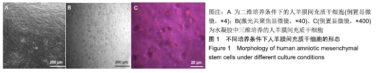
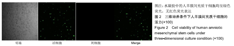
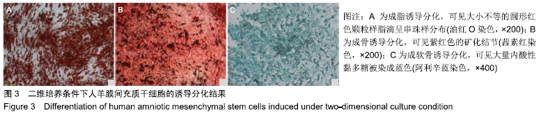
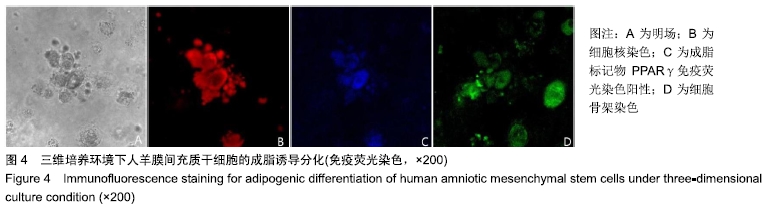
结果与结论:①二维培养组人羊膜间充质干细胞呈扁平状,为典型间充质样细胞形态;三维培养组人羊膜间充质干细胞呈圆形,均匀分散于水凝胶的每一层;②三维培养中的人羊膜间充质干细胞具有3系诱导分化潜能;③三维培养组白细胞介素6、白细胞介素8、表皮生长因子、碱性成纤维细胞因子、透明质酸、肝细胞生长因子、血管内皮生长因子mRNA相对表达量高于二维培养组(P < 0.001,P < 0.05),两组白细胞介素4、白细胞介素10、金属蛋白酶组织抑制因子、基质金属蛋白酶、转化生长因子、角化细胞生长因子mRNA相对表达量比较差异无显著性意义(P > 0.05);④三维培养组白细胞介素6、白细胞介素10、表皮生长因子、碱性成纤维细胞因子、白细胞介素8、肝细胞生长因子、转化生长因子β1、血管内皮生长因子蛋白表达量高于二维培养组(P < 0.001,P < 0.01,P < 0.05),两组白细胞介素4、金属蛋白酶组织抑制因子、基质金属蛋白酶、角化细胞生长因子蛋白表达量比较差异无显著性意义(P > 0.05);⑤结果表明,水凝胶三维培养环境中的人羊膜间充质干细胞可呈现更好的形态及创面修复相关因子旁分泌生物学效应。

ORCID: 0000-0002-9744-2176(王旗)

中国组织工程研究杂志出版内容重点:生物材料;骨生物材料; 口腔生物材料; 纳米材料; 缓释材料; 材料相容性;组织工程

关键词: 水凝胶, 羊膜间充质干细胞, 二维培养, 三维培养, 创面修复, 旁分泌效应

Abstract:

BACKGROUND: Studies have shown that mesenchymal stem cells can reduce inflammation, promote wound healing, and reduce scar formation in wound healing. However, previous two-dimensional culture environment can lead to differences in gene expression, signal transduction, and morphology because of intracellular contact inhibition. 

OBJECTIVE: To investigate whether the ability of wound healing related factors secreted by human amniotic mesenchymal stem cells is affected by two-dimensional or three-dimensional culture environment.

METHODS: The human amniotic mesenchymal stem cells cultured by traditional enzyme digestion method were inoculated in traditional cell culture flask (two-dimensional culture group) and ShakeGelTM 3D hydrogel (three-dimensional culture group) and induced to differentiate into adipocytes, osteoblasts, and chondrocytes, respectively. The direction of cell differentiation was determined by immunofluorescence staining. Human amniotic mesenchymal stem cells were fused to 70%-80% in two culture environments, and the growth characteristics and morphology of cells were observed under inverted phase contrast microscope and laser confocal microscope. After 24 hours of culture, relative mRNA expression of wound healing-related factors was detected by reverse transcription-quantitative polymerase chain reaction. After 48 hours of culture, the protein expression of wound healing-related factors was detected by the enzyme-linked immunosorbent assay.

RESULTS AND CONCLUSION: (1) Human amniotic mesenchymal stem cells cultured in two-dimensional culture group were flat and spindle-shaped, which was a typical mesenchymal stem cell-like morphology. Human amniotic mesenchymal stem cells in the three-dimensional culture group were round and evenly dispersed in each layer of the hydrogel. (2) Human amniotic mesenchymal stem cells in the three-dimensional culture group exhibited the potential to differentiate into adipocytes, osteoblasts, and chondrocytes. (3) The mRNA expression of interleukin-6, interleukin-8, epidermal growth factor, basic fibroblast growth factor, hyaluronic acid, hepatocyte growth factor, and vascular endothelial growth factor in the three-dimensional culture group was significantly higher than that in the two-dimensional culture group (P < 0.001, P < 0.05). There were no significant differences in the mRNA expression of interleukin-4, interleukin-10, tissue inhibitors of metalloproteinases, matrix metalloproteinase, transforming growth factor and keratinocyte growth factor between three-dimensional and two-dimensional culture groups (P > 0.05). (4) The protein expression of interleukin-6, interleukin-8, interleukin-10, epidermal growth factor, basic fibroblast growth factor, hepatocyte growth factor, transforming growth factor β1 and vascular endothelial growth factor in the three-dimensional culture group was significantly higher than that in the two-dimensional culture (P < 0.001, P < 0.01, P < 0.05). There were no significant differences in the protein expression of interleukin-4, tissue inhibitors of metalloproteinases, matrix metalloproteinase, and keratinocyte growth factor between two-dimensional and three-dimensional culture groups (P > 0.05). (5) These findings suggest that human amniotic mesenchymal stem cells cultured in three-dimensional hydrogel show better morphology and more encouraging paracrine effect of wound healing related factors than those cultured in traditional two-dimensional culture environment. 

Key words: hydrogel, amniotic mesenchymal stem cells, two-dimensional culture, three-dimensional culture, wound repair, paracrine effect

中图分类号: