中国组织工程研究 ›› 2013, Vol. 17 ›› Issue (32): 5749-5756.doi: 10.3969/j.issn.2095-4344.2013.32.002

• 骨髓干细胞 bone marrow stem cells • 上一篇    下一篇

骨髓间充质干细胞参与A549肺腺癌的组织修复

许  峰1,张  雷2,潘晋坤2,薛利利2,赵晓燕2,李宝平2   

  1. 1南通市肿瘤医院胸外科,江苏省南通市  226361
    2山西医科大学第二医院肿瘤生物科,山西省太原市  030001
  • 收稿日期:2012-12-21 修回日期:2013-02-26 出版日期:2013-08-06 发布日期:2013-08-06
  • 通讯作者: 李宝平,硕士,教授,主任医师,山西医科大学第二医院肿瘤生物科,山西省太原市 030001 164660904@qq.com
  • 作者简介:许峰★,男,1986年生,江苏省南通市人,汉族,2012年山西医科大学毕业,硕士,医师,主要从事胸部肿瘤的治疗与研究。 373599600@qq.com

Bone marrow mesenchymal stem cells are involved in tissue repair of A549 lung adenocarcinoma

Xu Feng1, Zhang Lei2, Pan Jin-kun2, Xue Li-li2, Zhao Xiao-yan2, Li Bao-ping2   

  1. 1Department of Chest Surgery, Nantong Tumor Hospital, Nantong  226361, Jiangsu Province, China
    2Department of Tumor Biological Treatment, the Second Hospital of Shanxi Medical University, Taiyuan  030001, Shanxi Province, China
  • Received:2012-12-21 Revised:2013-02-26 Online:2013-08-06 Published:2013-08-06
  • Contact: Li Bao-ping, Master, Professor, Chief physician, Department of Tumor Biological Treatment, the Second Hospital of Shanxi Medical University, Taiyuan 030001, Shanxi Province, China 164660904@qq.com
  • About author:Xu Feng★, Master, Physician, Department of Chest Surgery, Nantong Tumor Hospital, Nantong 226361, Jiangsu Province, China 373599600@qq.com

摘要:

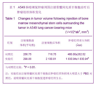
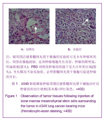
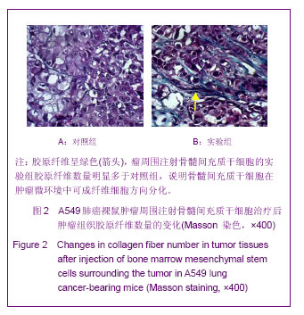
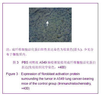
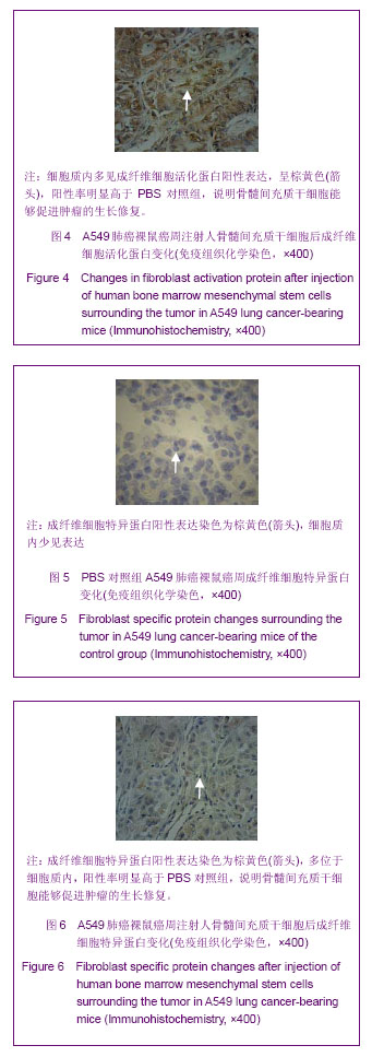
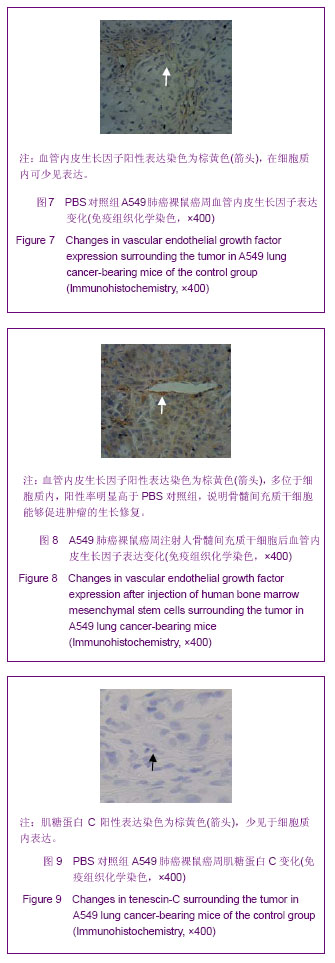
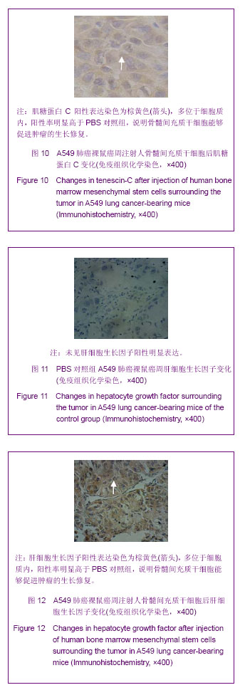
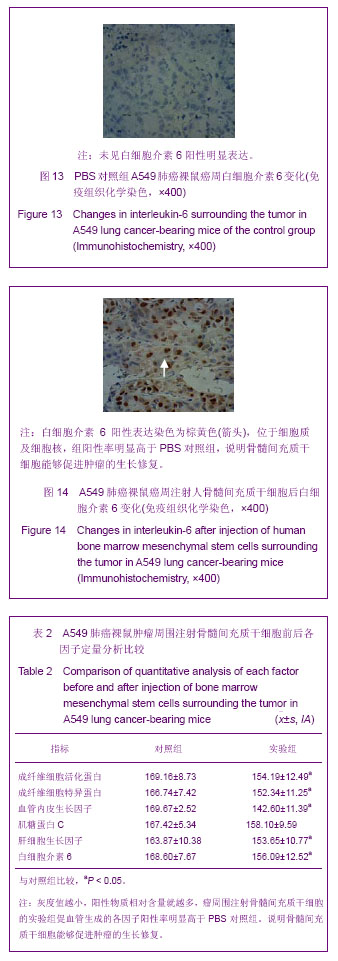
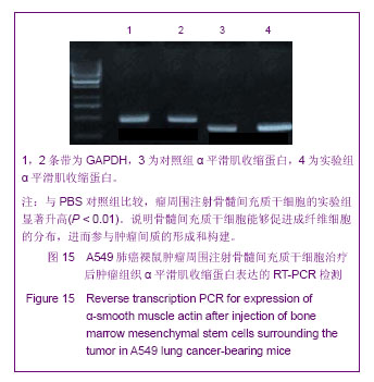
背景:肿瘤被认为是一种特殊的不愈合创伤,骨髓间充质干细胞通过向肿瘤组织归巢和向间质成分分化,参与肿瘤间质重构,从而改变肿瘤微环境,影响肿瘤的生长和转移。
目的:在A549肺癌荷瘤小鼠模型上证实骨髓间充质干细胞参与其损伤修复,探讨骨髓间充质干细胞参与肿瘤组织修复的机制。
方法:体外分离、培养人骨髓间充质干细胞,使用流式细胞术鉴定,制造A549肺癌荷瘤小鼠模型。实验组采用瘤周注射人骨髓间充质干细胞,对照组注射等量PBS,对比观察动物生活情况,肿瘤生长大小。4周后取材,苏木精-伊红染色对比观察肿瘤组织,Masson染色对比胶原纤维含量,RT-PCR检测两组α平滑肌收缩蛋白的表达,免疫组织化学检测两组成纤维细胞特异蛋白、成纤维细胞活化蛋白的表达情况,反映两组肿瘤组织的间质纤维的程度。免疫组织化学方法对比两组中血管内皮生长因子、肝细胞生长因子、白细胞介素6、肌糖蛋白C的表达高低。
结果与结论:骨髓间充质干细胞促进荷瘤小鼠肿瘤的生长,实验组肿瘤组织生长速度明显快于对照组(P < 0.05)。RT-PCR检测α平滑肌收缩蛋白的表达:与对照组比较,实验组α平滑肌收缩蛋白 mRNA的表达水平显著升高。免疫组织化学方法检测实验组肿瘤组织中TAFs标志物:成纤维细胞特异蛋白、成纤维细胞活化蛋白的表达,IHC检测血管内皮生长因子、肝细胞生长因子、白细胞介素6、肌糖蛋白C的表达明显高于对照组,差异有显著性意义(P < 0.05)。说明骨髓间充质干细胞在肿瘤微环境中向成纤维细胞方向分化,参与肿瘤间质的形成和构建,分泌血管内皮生长因子、肝细胞生长因子、白细胞介素6、肌糖蛋白C等促进肿瘤的生长修复。

关键词: 干细胞, 骨髓干细胞, 骨髓间充质干细胞, A549荷瘤小鼠肺癌, 肿瘤相关成纤维细胞, 间质纤维, 免疫组化, α平滑肌收缩蛋白, 血管内皮生长因子, 肝细胞生长因子, 白细胞介素6, 肌糖蛋白C, 干细胞图片文章

Abstract:

BACKGROUND: Tumor has been considered as a specific nonhealing trauma. Bone marrow mesenchymal stem cells participate in tumor mesenchymal reconstitution by tumor tissue homing and differentiation into mesenchyme, resulting in changing tumor microenvironment and affecting tumor growth and transfer.
OBJECTIVE: To explore the mechanisms of participation of bone marrow mesenchymal stem cells in tumor tissue repair in an A549 lung cancer-bearing mouse model.
METHODS: Bone marrow mesenchymal stem cells were isolated in vitro, cultured, and identified using flow cytometry, and then used to establish a mouse model of A549 lung cancer-bearing. In the experimental group, human bone marrow mesenchymal stem cells were injected into tissue surrounding the tumor. In the control group, an equal volume of PBS was injected. Animal survival condition and tumor size were compared. At 4 weeks, the specimens were harvested. Hematoxylin-eosin staining was used to compare tumor tissue. Masson staining was utilized to compare collagen fiber content. Reverse transcription-PCR was employed to detect the expression of α-smooth muscle actin. Immunohistochemistry was used to examine the expression of fibroblast specific protein and fibroblast activation protein to reflect the degree of interstitial fibers in tumor tissue in both groups. The expression levels of vascular endothelial growth factor, hepatocyte growth factor, interleukin-6 and tenescin-C were compared between the two groups using immunohistochemistry.
RESULTS AND CONCLUSION: Bone marrow mesenchymal stem cells promoted tumor growth in tumor-bearing mice. The growth rate of tumor tissue in experimental group was faster than the control group (P < 0.05). Compared with the control group, α-smooth muscle actin mRNA expression was significantly higher in the experimental group. Immunohistochemistry was used to detect the expression of tumor angiogenesis factors markers (fibroblast specific protein and fibroblast activation protein) in tumor tissue of experimental group. The expression levels of vascular endothelial growth factor, hepatocyte growth factor, interleukin-6 and tenescin-C were significantly greater in the experimental group than in the control group (P < 0.05). Results indicated that bone marrow mesenchymal stem cells differentiated into fibroblasts in tumor microenvironment, participated in the formation and construction of tumor stroma as well as promoted the growth and repair of tumor via the secretion of vascular endothelial growth factor, hepatocyte growth factor, interleukin-6 and tenescin-C.

Key words: stem cells, bone marrow-derived stem cells, bone marrow mesenchymal stem cells, A549 tumor-bearing mice, tumor-associated fibroblasts, stroma fiber, immunohistochemistry, α-smooth muscle actin, vascular endothelial growth factor, hepatocyte growth factor, interleukin-6, tenescin-C, stem cell photographs-containing paper

中图分类号: