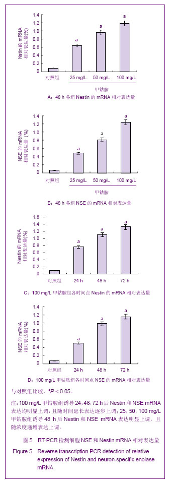
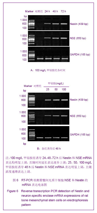
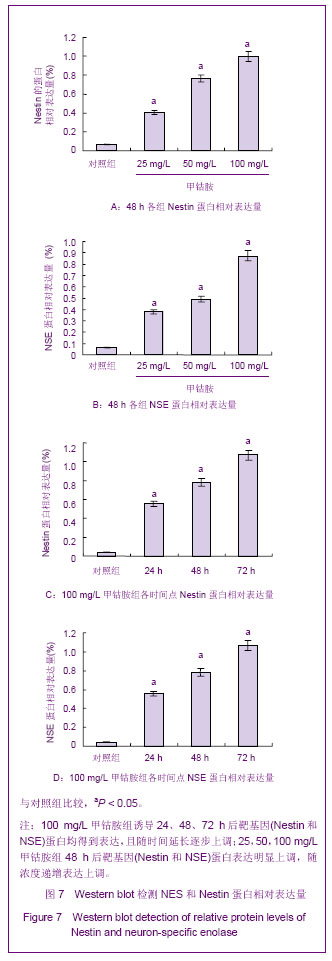
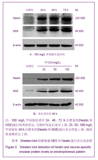
| [1] Delcroix GJ, Garbayo E, Sindji L, et al. Montero-Menei, the therapeutic potential of human multipotent mesenchymal stromal cells combined with pharmacologically active microcarriers transplanted in hemi-parkinsonian rats. Biomaterials. 2011;32(6):1560-1573.[2] Jiang J, Lv Z, Gu Y, et al. Adult rat mesenchymal stem cells differentiate into neuronal-like phenotype and express a variety of neuro-regulatory molecules in vitro. Neurosci Res. 2010;66(1):46-52.[3] Gnecchi M, Melo LG. Bone marrow-derived mesenchymal stem cells: isolation, expansion, characterization, viral transduction, and production of conditioned medium. Methods Mol Biol. 2009;482:281-294.[4] 张明鸣,贾贵清,罗婷,等.大鼠骨髓间充质干细胞体外两种分离方法和培养条件下生物学特点的比较[J].华西医学,2009,24(2): 371-373.[5] Pittenger MF, Mackay AM, Beck SC, et al. Multilineage potential of adult human mesenchymal stem cells. Science. 1999;284(5411):143-147.[6] Woodbury D, Schwarz EJ, Prockop DJ, et al. Adult rat and human bone marrow stromal cells differentiate into neurons. J Neurosci Res. 2000;61(4):364-370.[7] Li Q, Geng YJ, Lu L, et al. Platelet-rich fibrin-induced bone marrow mesenchymal stem cell differentiation into osteoblast-like cells and neural cells. Neural Regen Res. 2011;6(31):2419-2423.[8] Sanchez-Ramos J, Song S, Cardozo-Pelaez F, et al. Adult bone marrow stromal cells differentiate into neural cells in vitro. Exp Neurol. 2000;164(2):247-256.[9] 杜红阳,付海燕,包翠芬,等.地黄多糖对大鼠骨髓间充质干细胞向神经样细胞诱导分化作用的研究[J].中国实验方剂学杂志,2012, 18(6):133-137.[10] 辛颖,李玉林,张丽红.成人骨髓间充质干细胞体外定向诱导分化为神经元样细胞的研究[J].中国实验诊断学,2007,4(4):4-7.[11] 刘金保,董晓先,董燕湘,等.多种中药成分诱导大鼠骨髓间质干细胞转变为神经元样细胞[J].中国药物与临床,2003,3(3): 234-236.[12] Zhang Y, Wu C, Friis T, et al. The osteogenic properties of CaP/silk composite scaffolds. Biomaterials. 2010;31(10): 2848-2856.[13] Shevde N. Stem Cells: Flexible friends. Nature. 2012; 483 (7387):S22-26.[14] Liu Y, Zhang BA, Song Y, et al. Bone marrow mesenchymal stem cell transplantation for treatment of spinal cord injury: an in vivo magnetic resonance imaging tracking study. Neural Regen Res. 2011;6(13):978-982.[15] Ankeny DP, McTigue DM, Jakeman LB. Bone marrow transplants provide tissue protection and directional guidance for axons after contusive spinal cord injury in rats. Exp Neurol. 2004;190(1):17-31.[16] Wang J, Ding F, Gu Y, et al. Bone marrow mesenchymal stem cells promote cell proliferation and neurotrophic function of Schwann cells in vitro and in vivo. Brain Res. 2009;1262:7-15.[17] Wang D, Zhang JJ. Electrophysiological functional recovery in a rat model of spinal cord hemisection injury following bone marrow-derived mesenchymal stem cell transplantation under hypothermia. Neural Regen Res. 2012;7(10): 749-755.[18] Lee JB, Kuroda S, Shichinohe H, et al. A pre-clinical assessment model of rat autogeneic bone marrow stromal cell transplantation into the central nervous system. Brain Res Brain Res Protoc. 2004;14(1):37-44.[19] 朱旭萍,周智广.PGE1加甲钴胺治疗糖尿病周围神经病变[J].湖南医科大学学报,2001,26(4):343-344.[20] 张丽芳,杨晓苏,肖波,等.神经节苷脂对人骨髓间充质干细胞诱导分化为神经元样细胞生长状态的影响[J].脑与神经疾病杂志, 2009,17(1):13-15.[21] Vinores SA, Herman MM, Rubinstein LJ, et al. Electron microscopic localization of neuron-specific enolase in rat and mouse brain. J Histochem Cytochem. 1984;32(12): 1295-1302.[22] Lendahl U, Zimmerman LB, McKay RD. CNS stem cells express a new class of intermediate filament protein. Cell. 1990; 60(4):585-595.[23] Lothian C, Lendahl U. An evolutionarily conserved region in the second intron of the human nestin gene directs gene expression to CNS progenitor cells and to early neural crest cells. Eur J Neurosci. 1997;9(3):452-462.[24] Pekny M, Johansson CB, Eliasson C, et al. Abnormal reaction to central nervous system injury in mice lacking glial fibrillary acidic protein and vimentin. J Cell Biol. 1999;145(3):503-514. |
.jpg)
.jpg)