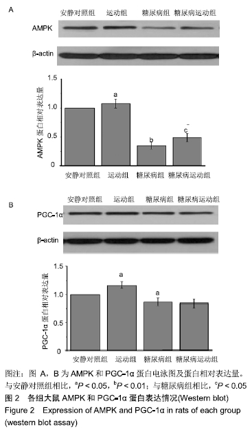
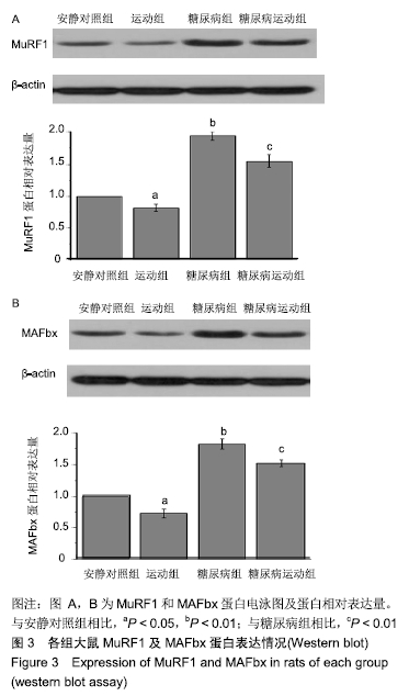
[1] 张坦,孙易,丁树哲.运动介导AMPK调控线粒体质量控制的机制研究进展[J].中国体育科技,2018,54(6):97-102.
[2] LIU HW, CHANG SJ. Moderate exercise suppresses NF-kappaB signaling and activates the SIRT1-AMPK-PGC1 alpha axis to attenuate muscle loss in diabetic db/db mice. Front Physiol. 2018; 636(9):1-9.
[3] BODINE SC, BAEHR LM. Skeletal muscle atrophy and the E3 ubiquitin ligases MuRF1 and MAFbx/atrogin-1. Am J Physiol Endocrinol Metab. 2014;307(6):469-484.
[4] LUNDBERG TR, FERNANDEZ-GONZALO R, TESCH PA, et al. Aerobic exercise augments muscle transcriptome profile of resistance exercise. Am J Physiol Regul Integr Comp Physiol. 2016;310(11): 1279-1287.
[5] KIM HJ, LEE WJ. Low-intensity aerobic exercise training: inhibition of skeletal muscle atrophy in high-fat-diet-induced ovariectomized rats. J Exerc Nutrition Biochem. 2017;21(3): 19-25.
[6] 高前进,柏海平,王彦伟.有氧运动对糖尿病大鼠自噬性肌萎缩的影响[J].中国康复医学杂志,2016,31(7):739-745.
[7] HUANG CC, WANG T, TUNG YT, et al. Effect of exercise training on skeletal muscle sirt1 and pgc-1alpha expression levels in rats of different age. Int J Med Sci. 2016;13(4): 260-270.
[8] LINDEN MA, FLETCHER JA, MORRIS EM, et al. Combining metformin and aerobic exercise training in the treatment of type 2 diabetes and NAFLD in OLETF rats. Am J Physiol Endocrinol Metab. 2014; 306(3): 300-310.
[9] ABD EL-KADER S, GARI A, SALAH EL-DEN A. Impact of moderate versus mild aerobic exercise training on inflammatory cytokines in obese type 2 diabetic patients: a randomized clinical trial. Afr Health Sci. 2013;13(4):857-863.
[10] KRAUSE M, RODRIGUES-KRAUSE J, O'HAGAN C, et al. The effects of aerobic exercise training at two different intensities in obesity and type 2 diabetes: implications for oxidative stress, low-grade inflammation and nitric oxide production. Eur J Appl Physiol. 2014;114(2):251-260.
[11] SRINIVASAN K, VISWANAD B, ASRAT L, et al. Combination of high-fat diet-fed and low-dose streptozotocin-treated rat: a model for type 2 diabetes and pharmacological screening. Pharmacol Res. 2005; 52(4):313-320.
[12] ARMSTRONG RB, OGILVIE RW, SCHWANE JA. Eccentric exercise- induced injury to rat skeletal muscle. J Appl Physiol Respir Environ Exerc Physiol. 1983;54(1):80-93.
[13] 王继,周越,张荷,等.Myostatin信号通路在4周离心耐力运动改善2型糖尿病大鼠骨骼肌萎缩中的作用[J].中国应用生理学杂志,2018, 34(3):223-228.
[14] 谭景旺,吴雪萍.全身振动训练对老年人下肢功能和慢性疾病影响的研究与进展[J].中国组织工程研究,2017,21(8):1288-1293.
[15] 程加秋,张庭然.低氧环境振动训练条件下2型糖尿病骨质疏松大鼠骨质代谢及胰岛素敏感性[J].中国组织工程研究,2018,22(12):1852-1858.
[16] ESPELAND MA, GLICK HA, BERTONI A, et al. Impact of an intensive lifestyle intervention on use and cost of medical services among overweight and obese adults with type 2 diabetes: the action for health in diabetes. Diabetes Care. 2014;37(9): 2548-2556.
[17] YANG Z, SCOTT CA, MAO C, et al. Resistance exercise versus aerobic exercise for type 2 diabetes: a systematic review and meta-analysis. Sports Med. 2014;44(4):487-499.
[18] MARTIN IK, KATZ A, WAHREN J. Splanchnic and muscle metabolism during exercise in NIDDM patients. Am J Physiol. 1995;269(3): 583-590.
[19] FOLLI F, SAAD MJ, BACKER JM, et al. Insulin stimulation of phosphatidylinositol 3-kinase activity and association with insulin receptor substrate 1 in liver and muscle of the intact rat. J Biol Chem. 1992;267(31):22171-22177.
[20] JOSEPH AM, ADHIHETTY PJ, LEEUWENBURGH C. Beneficial effects of exercise on age-related mitochondrial dysfunction and oxidative stress in skeletal muscle. J Physiol. 2016;594(18): 5105-5123.
[21] ROMANELLO V, SANDRI M. Mitochondrial quality control and muscle mass maintenance. Front Physiol. 2015;6:422.
[22] PLOMGAARD P, WEIGERT C. Do diabetes and obesity affect the metabolic response to exercise? Curr Opin Clin Nutr Metab Care. 2017;20(4):294-299.
[23] WANG M, LI S, WANG F, et al. Aerobic exercise regulates blood lipid and insulin resistance via the toll like receptor 4 mediated extracellular signal regulated kinases/AMP activated protein kinases signaling pathway. Mol Med Rep. 2018;17(6):8339-8348.
[24] JEON SM. Regulation and function of AMPK in physiology and diseases. Exp Mol Med. 2016;15;48(7):e245.
[25] KIM HJ, LEE WJ. Low-intensity aerobic exercise training: inhibition of skeletal muscle atrophy in high-fat-diet-induced ovariectomized rats. J Exerc Nutrition Biochem. 2017;21(3): 19-25.
[26] KAUPPINEN A, SUURONEN T, OJALA J, et al. Antagonistic crosstalk between NF-kappaB and SIRT1 in the regulation of inflammation and metabolic disorders.Cell Signal.2013;25(10): 1939-1948.
[27] 王继,周越.2型糖尿病与肌萎缩研究进展[J].中国运动医学杂志,2017, 36(7):645-650.
[28] HAWLEY JA. Molecular responses to strength and endurance training: are they incompatible? Appl Physiol Nutr Metab. 2009; 34(3):355-361.
[29] OSTLER JE, MAURYA SK, DIALS J, et al. Effects of insulin resistance on skeletal muscle growth and exercise capacity in type 2 diabetic mouse models. Am J Physiol Endocrinol Metab.2014; 306(6):592-605.
[30] AKAGAWA M, MIYAKOSHI N, KASUKAWA Y, et al. Effects of activated vitamin D, alfacalcidol, and low-intensity aerobic exercise on osteopenia and muscle atrophy in type 2 diabetes mellitus model rats. PLoS One. 2018;13(10):e0204857.
|
