中国组织工程研究 ›› 2019, Vol. 23 ›› Issue (18): 2806-2811.doi: 10.3969/j.issn.2095-4344.1732

• 组织工程骨及软骨材料 tissue-engineered bone and cartilage materials • 上一篇    下一篇

载重组人骨形态发生蛋白2/聚乳酸缓释微球的复合生物支架修复超临界骨缺损

周思睿,肖红利,黄文良,邓  江,叶  鹏
  

  1. 遵义医科大学第三附属医院骨科一病区,贵州省遵义市  563000
  • 收稿日期:2019-02-18 出版日期:2019-06-28 发布日期:2019-06-28
  • 通讯作者: 邓江,教授,主任医师,硕士生导师,遵义医科大学第三附属医院骨科一病区,贵州省遵义市 563000
  • 作者简介:周思睿,男,1984年生,汉族,重庆市人,硕士,主治医师,主要从事骨与关节损伤、运动医学研究。
  • 基金资助:

    义市科学技术局与遵义市第一人民医院联合科技研发资金项目[遵市科合社字(2018)162号],项目负责人:周思睿;国家自然科学基金(81660367),项目参与者:周思睿;贵州省科学技术基金[黔科合基础(2016)1420号],项目参与者:周思睿

Supercritical bone defect repaired by composite bioscaffolds loaded with recombinant human bone morphogenetic protein 2/poly(lactic acid)

Zhou Sirui, Xiao Hongli, Huang Wenliang, Deng Jiang, Ye Peng
  

  1. First Ward of Orthopedics, the Third Affiliated Hospital of Zunyi Medical University, Zunyi 563000, Guizhou Province, China
  • Received:2019-02-18 Online:2019-06-28 Published:2019-06-28
  • Contact: Deng Jiang, Professor, Chief physician, Master’s supervisor, First Ward of Orthopedics, the Third Affiliated Hospital of Zunyi Medical University, Zunyi 563000, Guizhou Province, China
  • About author:Zhou Sirui, Master, Attending physician, First Ward of Orthopedics, the Third Affiliated Hospital of Zunyi Medical University, Zunyi 563000, Guizhou Province, China
  • Supported by:

    the Zunyi Science and Technology Bureau and Zunyi First People’s Hospital Joint Science and Technology Research and Development Project, No. (2018)162 (to ZSR); the National Natural Science Foundation of China, No. 81660367 (to ZSR); the Science and Technology Foundation of Guizhou Province, No. (2016)1420 (to ZSR)

摘要:

文章快速阅读:


文题释义:
重组人骨形态发生蛋白2/聚乳酸缓释微球:重组人骨形态发生蛋白2是目前公认的修复骨缺损最重要的生长因子之一,与骨修复呈正相关;聚乳酸是可缓慢降解及生物相容性良好的生物高分子材料,作为缓释载体,具有良好的粒径及较稳定释药率,可使重组人骨形态发生蛋白2在骨骼缺损区持续释放,促进局部骨骼修复。
超临界骨缺损:骨骼缺损达到一定范围及距离后,机体自身无法自行修复,该临界点即为临界骨缺损。1986年Schmitz等基于动物实验模型提出了长骨干缺损达到长骨干直径的1.5倍即可认定为临界骨缺损,目前临床上将临界骨缺损的长度定义为1.5-3.0 cm。
 
 
背景:前期研究制备了丝素/壳聚糖/纳米羟基磷灰石复合生物支架与重组人骨形态发生蛋白2/聚乳酸缓释系统。
目的:观察载重组人骨形态发生蛋白2/聚乳酸缓释微球的复合生物支架,修复犬桡骨超临界骨缺损的效果。
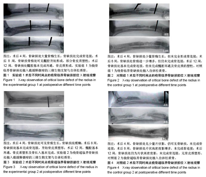
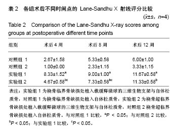
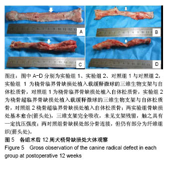
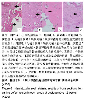
方法:采用真空冷冻干燥及化学交联法制备丝素蛋白/壳聚糖/纳米羟基磷灰石生物支架,采用复乳挥发法制备重组人骨形态发生蛋白2/聚乳酸缓释微球,将二者整合后制备成载缓释微球的三维生物支架。取8只成年犬(遵义医科大学实验动物中心提供),制作桡骨临界骨缺损模型(桡骨中段截骨3 cm),其中4只骨缺损处植入载缓释微球的三维生物支架与自体松质骨(实验组1),另4只仅植入自体松质骨(对照组1);另取8只成年犬,制作桡骨超临界骨缺损模型(桡骨中段截骨5 cm),其中4只骨缺损处植入载缓释微球的三维生物支架与自体松质骨(实验组2),另4只仅植入自体松质骨(对照组2)。术后 4,8,12周进行X射线检查;术后12周,取出缺损区桡骨,行组织病理学观察。实验已通过遵义医科大学伦理委员会审查,批准号:伦审(2016)2-071号。
结果与结论:①X射线:术后12周,实验组1缺损处髓腔基本再通,骨皮质形成,对照组1缺损处基本完成骨连接,但未完成髓腔再通与皮质骨塑形;实验组2髓腔基本再通,骨皮质塑形与骨长入基本完成,对照组2未完成骨连接,无皮质骨塑形;②组织学观察:实验组1骨缺损处已完成骨连接,可观察到骨皮质形成及完全髓腔再通,对照组1可见骨组织形成,其间可见大量纤维组织,无皮质骨,髓腔未完成再通;实验组2骨缺损处已有皮质骨形成及完成髓腔再通,对照组2可见少量骨组织,无皮质骨形成及髓腔再通;并且实验组1骨修复效果优于实验组2;③结果表明载重组人骨形态发生蛋白2缓释微球的丝素蛋白/壳聚糖/纳米羟基磷灰石三维生物支架,能有效修复一定范围内的犬桡骨临界及超临界骨缺损。

关键词: 骨形态发生蛋白2, 生物支架, 丝素蛋白, 壳聚糖, 纳米羟基磷灰石, 缓释微球, 超临界骨缺损, 自体松质骨

Abstract:

BACKGROUND: Preliminary study has prepared the silk fibroin/chitosan/nano-hydroxyapatite composite bioscaffold and the recombinant human bone morphogenetic protein 2/polylactic acid sustained release system.
OBJECTIVE: To observe the effect of radial bone defect repairing in dogs with composite bioscaffold loaded with recombinant human bone morphogenetic protein 2/poly(lactic acid) sustained-release microspheres.
METHODS: Silk fibroin/chitosan/nano-hydroxyphosphorous bioscaffolds were prepared by vacuum freeze-drying and chemical crosslinking. Recombinant human bone morphogenetic protein 2/poly(lactic acid) sustained-release microspheres were prepared by compound emulsion volatilization, and then prepared the three-dimensional bioscaffold. Eight adult dogs (provided by the Experimental Animal Center of Zunyi Medical University) were used to make a critical bone defect model of the radius (3 cm in the middle radius). Four bone defects were implanted with the three-dimensional bioscaffold with sustained release microspheres and autologous cancellous bones (experimental group 1). The other four only implanted with autologous cancellous bone (control group 1). Another eight adult dogs were used to make a superficial bone defect model of the radius (5 cm in the middle radius). Four bone defects were implanted with the three-dimensional biological scaffold with sustained release microspheres and autologous cancellous bone (experimental group 2). The other four were implanted only with autologous cancellous bone (control group 2). X-ray examination was performed at postoperative 4, 8, and 12 weeks. At postoperative 12 weeks, the radius of the defect area was removed for histopathological observation. The study was approved by the Ethics Committee of Zunyi Medical University, approval number: (2016)2-071.
RESULTS AND CONCLUSION: (1)X-ray: at postoperative 12 weeks, the medullary cavity in the experimental group 1 was recanalized and the cortical bone was formed. The defect in the control group 1 was basically completely connected, but the medullary cavity recanalization and cortical bone shaping were incomplete. In the experimental group 2, the medullary cavity was basically recanalized, the cortical bone shaping and bone ingrowth were basically completed. The control group 2 appeared no bone connection, no cortical bone shaping. (2) Histological observation: in the experimental group 1, the bone defect was connected completely, and the cortical bone formation and complete medullary cavity recanalization were observed. In the control group 1, bone tissue was formed, and a large amount of fibrous tissues were observed, no cortical bone was formed, and the medullary cavity was not recanalized. In experimental group 2, the cortical bone formation and the completion of the medullary cavity were recanalized in the bone defect. The control group 2 showed a small amount of bones, no cortical bone formation and recanalization of the medullary cavity. The experimental group 1 has better bone repair effect than the experimental group 2. (3) These results imply that the three-dimensional bioscaffolds loaded with recombinant human bone morphogenetic protein 2/poly (lactic acid) sustained-release microspheres can effectively repair the critical and supercritical bone defects of canine tibia to some extent.

Key words: bone morphogenetic protein-2, bioscaffold, silk fibroin, chitosan, nano-hydroxyapatite, sustained release microspheres, supercritical bone defect;, autologous cancellous bone

中图分类号: