中国组织工程研究 ›› 2021, Vol. 25 ›› Issue (10): 1555-1559.doi: 10.3969/j.issn.2095-4344.3031

• 药物控释材料 drug delivery materials • 上一篇    下一篇

几丁糖/聚乙烯醇导管联合脑源性神经营养因子缓释微球修复大鼠周围神经缺损

史正亮,张  华,范志勇,马  维,袁  鹤,杨  冰   

  1. 河北医科大学第二医院骨科,河北省石家庄市   050000
  • 收稿日期:2020-03-21 修回日期:2020-03-26 接受日期:2020-05-07 出版日期:2021-04-08 发布日期:2020-12-18
  • 通讯作者: 张华,副主任医师,河北医科大学第二医院骨科,河北省石家庄市 050000
  • 作者简介:史正亮,男,1977年生,河北省石家庄市人,汉族,1999年天津医科大学毕业,硕士,副主任医师,主要从事脊柱及周围神经损伤方面的研究。

Nerve conduits of chitosan/polyvinyl alcohol with brain-derived neurotrophic factor microspheres for peripheral nerve defects in rats

Shi Zhengliang, Zhang Hua, Fan Zhiyong, Ma Wei, Yuan He, Yang Bing   

  1. Department of Orthopedics, the Second Hospital of Hebei Medical University, Shijiazhuang 050000, Hebei Province, China
  • Received:2020-03-21 Revised:2020-03-26 Accepted:2020-05-07 Online:2021-04-08 Published:2020-12-18
  • Contact: Zhang Hua, Associate chief physician, Department of Orthopedics, the Second Hospital of Hebei Medical University, Shijiazhuang 050000, Hebei Province, China
  • About author:Shi Zhengliang, Master, Associate chief physician, Department of Orthopedics, the Second Hospital of Hebei Medical University, Shijiazhuang 050000, Hebei Province, China

摘要:

文题释义:
生物微囊:是通过将酶、蛋白质和激素等生物活性物质包封在选择性透过膜中,形成球状微胶囊,通过微囊膜的选择透过作用发挥良好的缓释、控释和靶向递药作用。
神经导管支架:神经导管支架材料分为天然材料和合成材料,合成材料因可以得到可控的化学、物理特性,是常用的支架材料,其中生物可降解材料是目前研究的热点。神经导管支架修复神经缺损一方面可以提供保证神经再生的通道,防止周围结缔组织侵入和瘢痕的形成;另一方面,远侧神经支产生的趋化性营养因子和加入的外源性神经营养因子、生长因子等能在导管内积聚,对再生轴突的生长方向产生重要影响。 
脑源性神经营养因子:是神经营养家族的重要成员之一,主要分布于脑,在心、肺和脊髓运动神经元的外周靶组织也有一定的分布,对神经元的存活、分化、生长发育起重要作用,而且能促进受损伤神经元再生及分化等生物效应,是中枢及周围神经系统的重要营养因子。

背景:通过神经导管桥接修复周围神经缺损可为神经再生提供适合的微环境,一方面为神经再生提供唯一的通道,防止周围结缔组织的侵入和瘢痕的形成;另一方面,能保存内源性和外源性的神经营养因子、生长因子等促进轴突生长的刺激物质。
目的:观察几丁糖/聚乙烯醇导管内注入脑源性神经营养因子缓释微球桥接周围神经缺损的效果。
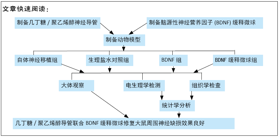
方法:采用反复冻融技术制备几丁糖/聚乙烯醇神经导管;采用乳化-溶剂挥发法与聚合物合金法制备脑源性神经营养因子缓释微球。在60只成年雄性SD大鼠右后肢制作15 mm坐骨神经缺损模型,随机分4组处理:A组植入自体坐骨神经,B组植入几丁糖/聚乙烯醇神经导管后注射生理盐水,C组植入几丁糖/聚乙烯醇神经导管后注射脑源性神经营养因子溶液,D组植入几丁糖/聚乙烯醇神经导管后注射脑源性神经营养因子缓释微球,每组15只。术后4个月取材,进行大体解剖观察、组织学检查和神经电生理测定。动物实验获得河北医科大学第二医院科研伦理委员会批准。
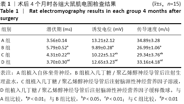
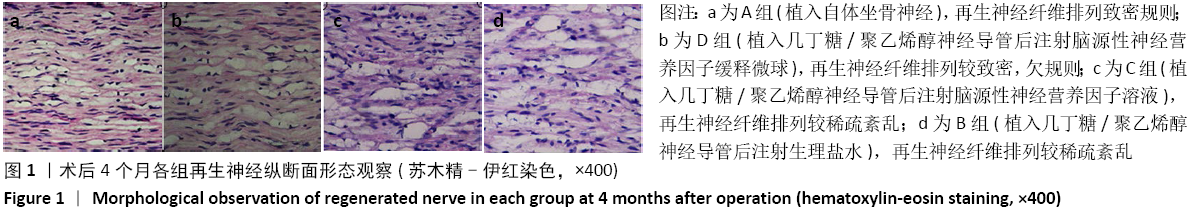
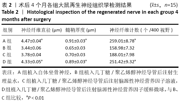
结果与结论:①大体解剖:A、D组肌肉萎缩较另外2组轻,4组移植物均与周边组织有粘连,自体移植段神经与周围组织粘连较重,D组再生神经已将远、近两端神经连接,再生神经填满导管;②电生理检测:D组潜伏期短于B、C组(P < 0.01),诱发电位与传导速度大于B、C组(P < 0.01),与A组比较差异均无显著性意义(P > 0.05);③组织学观察:B、C、D组导管内均有再生神经纤维,D组再生神经纤维直径、个数及髓鞘厚度大于B、C组(P < 0.01),与A组比较差异均无显著性意义(P > 0.05);④结果表明,几丁糖/聚乙烯醇导管内注入脑源性神经营养因子微球对于外周神经再生有较长期的促进作用。

https://orcid.org/0000-0002-2815-6109 (史正亮) 
中国组织工程研究杂志出版内容重点:生物材料;骨生物材料; 口腔生物材料; 纳米材料; 缓释材料; 材料相容性;组织工程

关键词: 材料, 支架, 神经, 神经导管, 周围神经, 神经缺损, 脑源性神经营养因子, 缓释微球, 神经再生

Abstract: BACKGROUND:  The repair of peripheral nerve defects by nerve conduit bridging can provide a suitable microenvironment for nerve regeneration. On one hand, it can provide a unique channel for nerve regeneration, prevent the invasion of peripheral connective tissue and the formation of scars. On the other hand, it can maintain endogenous and exogenous neurotrophic factors, growth factors and other stimulants to promote axon growth.
OBJECTIVE: To observe the therapeutic effect of chitosan/polyvinyl alcohol catheter injected with brain-derived neurotrophic factor sustained-release microspheres to bridge peripheral nerve defects.
METHODS: Chitosan/polyvinyl alcohol nerve conduit was prepared by repeated freeze-thaw technique. The brain-derived neurotrophic factor microspheres were obtained by polymer-alloys combined with oil-oil emulsion/solvent evaporation method. A 15 mm sciatic nerve defect model was made in the right hindlimb of 60 adult male Sprague-Dawley rats. They were selected and randomly divided into four groups (n=15 per group): group A implanted with autogenous sciatic nerve; group B implanted with chitosan/polyvinyl alcohol nerve catheter, injected with normal saline; group C implanted with chitosan/polyvinyl alcohol nerve catheter, injected with brain-derived neurotrophic factor solution; group D implanted with chitosan/polyvinyl alcohol nerve catheter, injected with brain-derived neurotrophic factor sustained-release microspheres. General observation, histological inspection, and electrophysiological determination were performed at 4 months after the surgery. This study was approved by the Research Ethics Committee of the Second Hospital of Hebei Medical University. 
RESULTS AND CONCLUSION: (1) Gross anatomy showed that muscle atrophy in group A and group D was lighter than that in the other two groups. The grafts in four groups were all adhered to the peripheral tissues, and the nerve in the autotransplantation segment was strongly adhered to the peripheral tissues. In group D, the regenerated nerve had connected the distal and proximal nerves, and the regenerated nerve filled the conduit. (2) Electrophysiological examination showed that the latency of group D was shorter than that of groups B and C (P < 0.01), and the evoked potential and conduction velocity of group D were higher than that of groups B and C (P  < 0.01). There was no significant difference between group D and group A (P > 0.05). (3) Histological observation showed that there were regenerated nerve fibers in groups B, C, and D. The diameter, number and thickness of myelin sheath of group D were larger than those of group B and group C (P < 0.01). There was no significant difference between group D and group A (P > 0.05). (4) The results showed that the injection of brain-derived neurotrophic factor microspheres into chitosan/PVA catheter had a long-term promoting effect on peripheral nerve regeneration.


Key words: materials, scaffold, nerve, nerve conduit, peripheral nerve, nerve defect, brain-derived neurotrophic factor, sustained release microspheres, nerve regeneration

中图分类号: