中国组织工程研究 ›› 2019, Vol. 23 ›› Issue (19): 3062-3066.doi: 10.3969/j.issn.2095-4344.1116
• 组织构建实验造模 experimental modeling in tissue construction • 上一篇 下一篇
基于模型迭代重建算法评估其对肺气肿区域的定量分析能力
张锦惠1,钟诗雨2
- (1成都医学院,四川省成都市 610500;2成都医学院第一附属医院,四川省成都市 610500 )
Pulmonary emphysema quantification on ultra-low-dose CT using model-based iterative reconstruction
Zhang Jinhui1, Zhong Shiyu2
- (1Chengdu Medical College, Chengdu 610500, Sichuan Province, China; 2the First Affiliated Hospital of Chengdu Medical College, Chengdu 610500, Sichuan Province, China)
摘要:
文章快速阅读:
.jpg) 文题释义:
迭代重建算法(Iterative Reconstruction,IR):是近年来新研发的一种图像重建算法,由于在计算中引入统计重建方法,具有可在数据不完全和低信噪比(低剂量)条件下成像的特点,从而克服了传统滤过反投影法算法在低剂量扫描时的缺点,使CT仅需极少的采样数据即可完成高质量成像,由此带来辐射剂量的大幅降低。
定量分析:定量分析起源于分析化学的一个分支。测定物质中各成分的含量使用方法不同,可分重量分析、容量分析和仪器分析3类。因分析试样用量和被测成分不同,又可分为常量分析、半微量分析、微量分析、超微量分析和痕量分析等。 指分析一个被研究对象所包含成分的数量关系或所具备性质间的数量关系;也可以对几个对象的某些性质、特征、相互关系从数量上进行分析比较,研究的结果也用“数量”加以描述。
文题释义:
迭代重建算法(Iterative Reconstruction,IR):是近年来新研发的一种图像重建算法,由于在计算中引入统计重建方法,具有可在数据不完全和低信噪比(低剂量)条件下成像的特点,从而克服了传统滤过反投影法算法在低剂量扫描时的缺点,使CT仅需极少的采样数据即可完成高质量成像,由此带来辐射剂量的大幅降低。
定量分析:定量分析起源于分析化学的一个分支。测定物质中各成分的含量使用方法不同,可分重量分析、容量分析和仪器分析3类。因分析试样用量和被测成分不同,又可分为常量分析、半微量分析、微量分析、超微量分析和痕量分析等。 指分析一个被研究对象所包含成分的数量关系或所具备性质间的数量关系;也可以对几个对象的某些性质、特征、相互关系从数量上进行分析比较,研究的结果也用“数量”加以描述。摘要
背景:随着设备的改进和算法的优化,最新一代搭配肺部参数设置的模型迭代重建算法可以提供针对肺部组织的更高分辨率图像。
目的:采用基于模型的迭代重建算法重建超低剂量CT的图像,并通过与标准剂量CT的图像进行对比,评估其对肺气肿区域的定量分析能力。
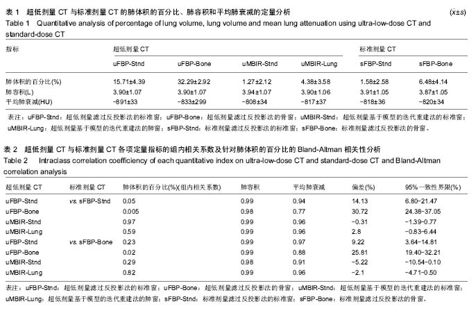
方法:回顾性地分析了45例接受肺部超低剂量CT和标准剂量CT(6.66±2.69)mSv扫描的患者。采用平滑滤波设置和锐化滤波设置的滤过反投影算法以及具有常规参数设置和肺参数设置的模型迭代重建算法重建CT图像,并且记录患者2次CT扫描的辐射剂量。以肺体积的百分比来评估患者的肺气肿程度,通过组内相关系数和Bland-Altman分析评估超低剂量CT和标准剂量CT之间的相关性。
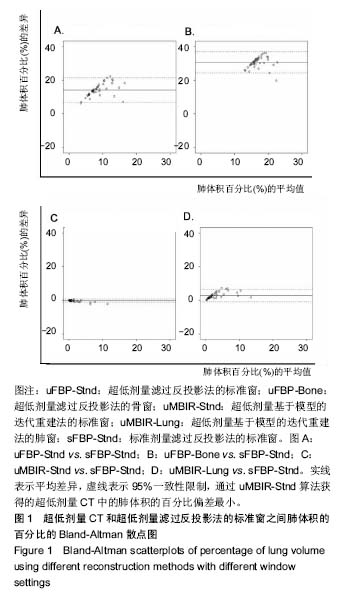
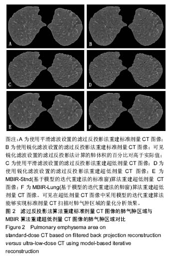
结果与结论:①超低剂量CT扫描的辐射剂量为(0.18±0.02)mSv,显著低于标准剂量CT。以平滑滤波设置的滤过反投影法算法重建的标准剂量CT图像作为参考标准,发现MBIR-Lung算法重建的超低剂量CT的相关性最好(组内相关系数=0.97,偏倚=-0.31%);②以锐化滤波设置的滤过反投影法算法重建的标准剂量CT作为参考标准,发现MBIR-Stnd算法重建的超低剂量CT的相关性较好(组内相关系数=0.82,偏差= -2.10%);③结果说明,基于模型的迭代重建算法能够显著提高超低剂量CT和标准剂量CT之间针对肺气肿量化分析的一致性。
中图分类号:
.jpg) 文题释义:
迭代重建算法(Iterative Reconstruction,IR):是近年来新研发的一种图像重建算法,由于在计算中引入统计重建方法,具有可在数据不完全和低信噪比(低剂量)条件下成像的特点,从而克服了传统滤过反投影法算法在低剂量扫描时的缺点,使CT仅需极少的采样数据即可完成高质量成像,由此带来辐射剂量的大幅降低。
定量分析:定量分析起源于分析化学的一个分支。测定物质中各成分的含量使用方法不同,可分重量分析、容量分析和仪器分析3类。因分析试样用量和被测成分不同,又可分为常量分析、半微量分析、微量分析、超微量分析和痕量分析等。 指分析一个被研究对象所包含成分的数量关系或所具备性质间的数量关系;也可以对几个对象的某些性质、特征、相互关系从数量上进行分析比较,研究的结果也用“数量”加以描述。
文题释义:
迭代重建算法(Iterative Reconstruction,IR):是近年来新研发的一种图像重建算法,由于在计算中引入统计重建方法,具有可在数据不完全和低信噪比(低剂量)条件下成像的特点,从而克服了传统滤过反投影法算法在低剂量扫描时的缺点,使CT仅需极少的采样数据即可完成高质量成像,由此带来辐射剂量的大幅降低。
定量分析:定量分析起源于分析化学的一个分支。测定物质中各成分的含量使用方法不同,可分重量分析、容量分析和仪器分析3类。因分析试样用量和被测成分不同,又可分为常量分析、半微量分析、微量分析、超微量分析和痕量分析等。 指分析一个被研究对象所包含成分的数量关系或所具备性质间的数量关系;也可以对几个对象的某些性质、特征、相互关系从数量上进行分析比较,研究的结果也用“数量”加以描述。