中国组织工程研究 ›› 2019, Vol. 23 ›› Issue (3): 378-383.doi: 10.3969/j.issn.2095-4344.1024
• 血管组织构建 vascular tissue construction • 上一篇 下一篇
血管内皮生长因子联合突变型低氧诱导因子1α的促血管生成作用
胡 亮1,王军海1,王志烈1,谢金元1,陈 登1,丁 凡2
- (荆门市第一人民医院,1关节外科,2创伤手足外科,湖北省荆门市 448000)
Vascular endothelial growth factor combined with mutant hypoxia-inducible factor 1alpha promotes angiogenesis
Hu Liang1, Wang Junhai1, Wang Zhilie1, Xie Jinyuan1, Chen Deng1, Ding Fan2
- (1Department of Joint Surgery, 2Department of Traumatic Hand and Foot Surgery, Jingmen No.1 People’s Hospital, Jingmen 448000, Hubei Province, China)
摘要:
文章快速阅读:
.jpg) 文题释义:
内皮祖细胞(endothelial progenitor cells,EPCs):是血管内皮细胞的前体细胞,亦称为成血管细胞(angioblast),在生理或病理因素刺激下,可从骨髓动员到外周血参与损伤血管的修复。1997年,Asahara等首次证明循环外周血中存在能分化为血管内皮细胞的前体细胞,并将其命名为血管内皮祖细胞。
微血管密度:是指生物组织如皮肤、肌肉、器官等组织中单位密度的微血管数量,在微循环研究中常把单位面积(1 mm2或某一长度范围内)所看到的血管数称为血管密度。在组织标本中可以在显微镜下计数单位面积的毛细血管数或单位体积的毛细血管数,微血管包括细动脉、细静脉、毛细血管。
文题释义:
内皮祖细胞(endothelial progenitor cells,EPCs):是血管内皮细胞的前体细胞,亦称为成血管细胞(angioblast),在生理或病理因素刺激下,可从骨髓动员到外周血参与损伤血管的修复。1997年,Asahara等首次证明循环外周血中存在能分化为血管内皮细胞的前体细胞,并将其命名为血管内皮祖细胞。
微血管密度:是指生物组织如皮肤、肌肉、器官等组织中单位密度的微血管数量,在微循环研究中常把单位面积(1 mm2或某一长度范围内)所看到的血管数称为血管密度。在组织标本中可以在显微镜下计数单位面积的毛细血管数或单位体积的毛细血管数,微血管包括细动脉、细静脉、毛细血管。摘要
背景:研究表明,血管内皮生长因子的转录翻译过程在许多缺血、缺氧的条件下都会增加,有效改善机体血管形成与侧支微循环。突变低氧诱导因子1α的表达也需要严格的缺氧条件,很大程度上限制了其实际应用。
目的:观察腺病毒介导的血管内皮生长因子联合突变型低氧诱导因子1α(Ad-VEGF-IRES-HIF-1αmu)转染内皮祖细胞在激素性股骨头缺血性坏死修复中促血管生成的作用。
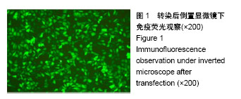
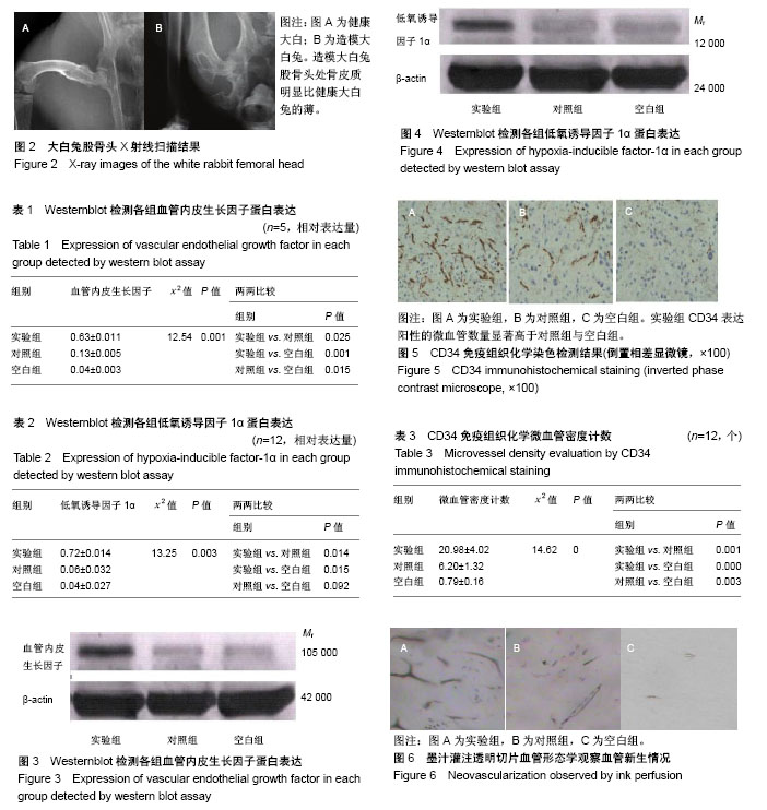
方法:①转染Ad-VEGF-IRES-HIF-1αmu到内皮祖细胞,观察细胞活性、形态及细胞病变效应;②将转染Ad-VEGF-IRES-HIF-1αmu成功的内皮祖细胞植入激素性股骨头缺血性坏死动物模型的股骨坏死部位(实验组:转染Ad-VEGF-IRES-HIF-1αmu,对照组:内皮祖细胞细胞悬液,空白组:细胞培养液);③移植10周后,检测血管内皮生长因子、低氧诱导因子1α蛋白表达及CD34表达和微血管密度计数;④墨汁灌注透明切片血管形态学观察。
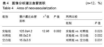
结果与结论:①实验组血管内皮生长因子和低氧诱导因子1α蛋白表达高于对照组与空白组(P < 0.05);②实验组的CD34表达阳性的微血管数量较多,且相互之间有连接。③实验组动物股骨头血管墨汁染色显示有新的血管生成,部分血管管径良好,有再通现象,血管间有清晰的连通脉络,有效的血管脉络均匀分布在缺损区域;实验组新生血管面积显著高于对照组和空白组(P < 0.05);④结果提示,血管内皮生长因子联合突变型低氧诱导因子1α在激素性股骨头缺血性坏死修复中可以增强血管生成作用。
中国组织工程研究杂志出版内容重点:组织构建;骨细胞;软骨细胞;细胞培养;成纤维细胞;血管内皮细胞;骨质疏松;组织工程
ORCID: 0000-0003-4580-4953(胡亮)
中图分类号:
.jpg) 文题释义:
内皮祖细胞(endothelial progenitor cells,EPCs):是血管内皮细胞的前体细胞,亦称为成血管细胞(angioblast),在生理或病理因素刺激下,可从骨髓动员到外周血参与损伤血管的修复。1997年,Asahara等首次证明循环外周血中存在能分化为血管内皮细胞的前体细胞,并将其命名为血管内皮祖细胞。
微血管密度:是指生物组织如皮肤、肌肉、器官等组织中单位密度的微血管数量,在微循环研究中常把单位面积(1 mm2或某一长度范围内)所看到的血管数称为血管密度。在组织标本中可以在显微镜下计数单位面积的毛细血管数或单位体积的毛细血管数,微血管包括细动脉、细静脉、毛细血管。
文题释义:
内皮祖细胞(endothelial progenitor cells,EPCs):是血管内皮细胞的前体细胞,亦称为成血管细胞(angioblast),在生理或病理因素刺激下,可从骨髓动员到外周血参与损伤血管的修复。1997年,Asahara等首次证明循环外周血中存在能分化为血管内皮细胞的前体细胞,并将其命名为血管内皮祖细胞。
微血管密度:是指生物组织如皮肤、肌肉、器官等组织中单位密度的微血管数量,在微循环研究中常把单位面积(1 mm2或某一长度范围内)所看到的血管数称为血管密度。在组织标本中可以在显微镜下计数单位面积的毛细血管数或单位体积的毛细血管数,微血管包括细动脉、细静脉、毛细血管。