中国组织工程研究 ›› 2020, Vol. 24 ›› Issue (25): 4006-4011.doi: 10.3969/j.issn.2095-4344.2095

• 牙髓及牙周膜干细胞 Dental pulp and periodontal ligament stem cells • 上一篇    下一篇

前列腺素E1联合碱性成纤维细胞生长因子干预人牙髓干细胞增殖和血管生成能力的变化

项海东,程东梅,郭  晗,高  琪   

  1. 河北医科大学第二医院口腔内科,河北省石家庄市  050000

  • 收稿日期:2019-09-07 修回日期:2019-09-09 接受日期:2019-10-31 出版日期:2020-09-08 发布日期:2020-08-24
  • 通讯作者: 高琪,副主任医师,副教授,硕士生导师,河北医科大学第二医院口腔内科,河北省石家庄市 050000
  • 作者简介:项海东,男,1982年生,硕士,主治医师,主要从事口腔牙体牙髓病方面的研究。

Changes in the proliferation and angiogenesis of human dental pulp stem cells after treated with prostaglandin E1 combined with basic fibroblast growth factor  

Xiang Haidong, Cheng Dongmei, Guo Han, Gao Qi    

  1. Department of Stomatology, the Second Hospital of Hebei Medical University, Shijiazhuang 050000, Hebei Province, China

  • Received:2019-09-07 Revised:2019-09-09 Accepted:2019-10-31 Online:2020-09-08 Published:2020-08-24
  • Contact: Gao Qi, Associate chief physician, Associate professor, Master’s supervisor, Department of Stomatology, the Second Hospital of Hebei Medical University, Shijiazhuang 050000, Hebei Province, China
  • About author:Xiang Haidong, Master, Attending physician, Department of Stomatology, the Second Hospital of Hebei Medical University, Shijiazhuang 050000, Hebei Province, China

摘要:


文题释义:

前列腺素E1是一种生物活性物质,广泛存在于体内各组织细胞,由BERGSRTOEM等于1960年首先分离获得,并详细研究了其分子结构,具有舒张血管、稳定细胞膜降低血小板黏附率、改善血液黏度及抗炎等作用,可促进骨髓间充质干细胞和神经干细胞的增殖、迁移,并促进细胞分泌血管内皮生长因子,进而促进血管生成。

碱性成纤维细胞生长因子:是一种肝素黏合多肽,也是一种重要的潜在有丝分裂原,属于成纤维细胞生长因子家族,对细胞有丝分裂及分化具有很强的调节作用,特别是对中胚层和神经外胚层来源的细胞作用更强,可促进细胞生长,研究发现牙髓中的成纤维细胞可表达碱性成纤维细胞生长因子,在体外能明显促进人牙髓干细胞增殖。


摘要

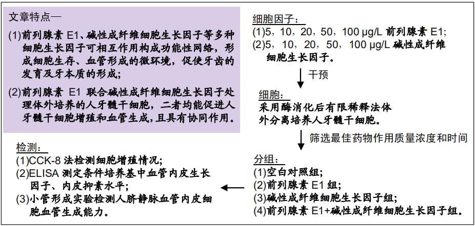
背景:前列腺素E1及碱性成纤维细胞生长因子可促使人牙髓干细胞增殖,但两者联合应用对人牙髓干细胞增殖和血管生成能力的影响还未见研究。

目的:探究前列腺素E1联合碱性成纤维细胞生长因子对人牙髓干细胞增殖和血管生成能力的影响。
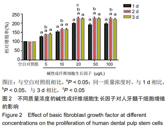
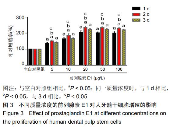
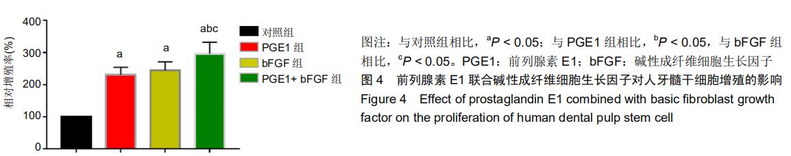
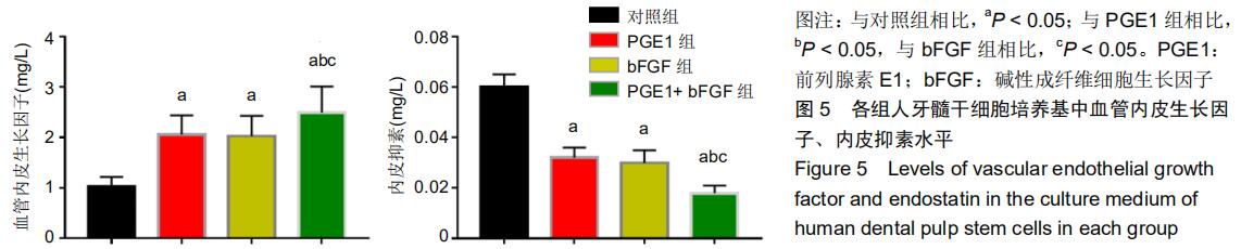
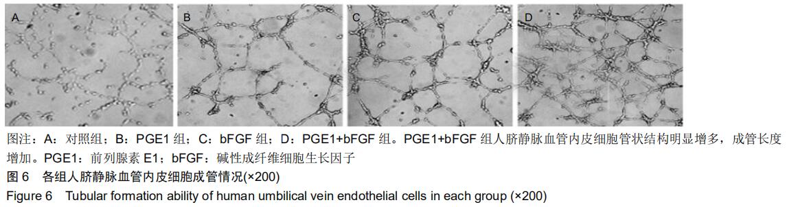
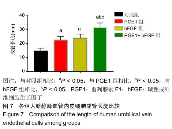
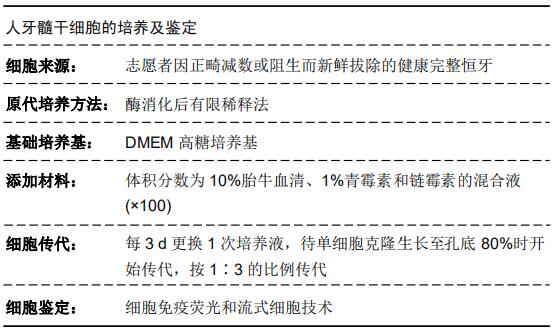
方法:体外分离培养人牙髓干细胞,经表面标志物检测鉴定后,分别以5102050100 μg/L前列腺素E15102050100 μg/L碱性成纤维细胞生长因子单独作用于体外培养的人牙髓干细胞,以未加药物处理的细胞作为空白对照组,采用CCK-8法分别在123 d检测人牙髓干细胞增殖情况,筛选最佳药物作用质量浓度和时间。将体外培养的人牙髓干细胞分为4组:空白对照组、前列腺素E1组、碱性成纤维细胞生长因子组、前列腺素E1+碱性成纤维细胞生长因子组,采用CCK-8法检测人牙髓干细胞增殖情况,然后提取人牙髓干细胞条件培养基,以ELISA测定条件培养基中血管内皮生长因子、内皮抑素水平,以小管形成实验检测人牙髓干细胞条件培养基干预后人脐静脉血管内皮细胞的体外小管形成能力。

结果与结论:前列腺素E1、碱性成纤维细胞生长因子的最佳作用质量浓度均为20 μg/L,最佳作用时间为2 d与空白对照组相比,前列腺素E1组、碱性成纤维细胞生长因子组、前列腺素E1+碱性成纤维细胞生长因子组人牙髓干细胞相对增殖率、血管内皮生长因子水平、人脐静脉血管内皮细胞体外血管生成能力均显著升高(P < 0.05),内皮抑素水平显著降低(P < 0.05);前列腺素E1+碱性成纤维细胞生长因子组上述各指标均优于前列腺素E1组、碱性成纤维细胞生长因子组(P < 0.05)结果表明,前列腺素E1联合碱性成纤维细胞生长因子可促进人牙髓干细胞增殖,增强人脐静脉血管内皮细胞体外小管形成能力。

 ORCID: 0000-0002-7727-1883(项海东)

中国组织工程研究杂志出版内容重点:干细胞;骨髓干细胞;造血干细胞;脂肪干细胞;肿瘤干细胞;胚胎干细胞;脐带脐血干细胞;干细胞诱导;干细胞分化;组织工程


关键词:

前列腺素E1, 碱性成纤维细胞生长因子,  人牙髓干细胞,  细胞增殖,  血管生成

Abstract:

BACKGROUND: Prostaglandin E1 and basic fibroblast growth factor can promote the proliferation of human dental pulp stem cells, but the effects of their combinations on the proliferation of human dental pulp stem cells and angiogenesis are unknown.

OBJECTIVE: To investigate the effects of prostaglandin E1 combined with basic fibroblast growth factor on the proliferation and angiogenesis of human dental pulp stem cells.

METHODS: (1) Human dental pulp stem cells were isolated and cultured in vitro. After detection and identification of surface markers, prostaglandin E1 and basic fibroblast growth factor at concentrations of 5, 10, 20, 50 and 100 μg/L were used to treat human dental pulp stem cells in vitro. The untreated cells served as control group. The cell proliferation was detected by cell counting kit-8 assay, and the optimum drug concentration and time of drug action were screened. (2) The in vitro cultured human dental pulp stem cells were divided into four groups: blank control group, prostaglandin E1 group, basic fibroblast growth factor group and combination group. The cell proliferation was detected by cell counting kit-8 assay. Human dental pulp stem cell conditioned medium was extracted. The levels of vascular endothelial growth factor and endostatin in the culture medium were detected by ELISA. The in vitro tubular formation ability of human umbilical vein endothelial cells after culture in conditioned medium was tested by tubule formation experiment.

RESULTS AND CONCLUSION: The optimum concentration of prostaglandin E1 and basic fibroblast growth factor was 20 μg/L, and the optimum time of action was 2 days. Compared with the blank control group, the relative proliferation rate, level of vascular endothelial growth factor and the angiogenesis ability of human umbilical vein endothelial cell in vitro in the prostaglandin E1, basic fibroblast growth factor and combination groups were significantly increased (P < 0.05), while the level of endostatin was significantly decreased (P < 0.05). All above index levels in the combination group were significantly superior to those in the prostaglandin E1 and basic fibroblast growth factor groups

(P < 0.05). In summary, prostaglandin E1 combined with basic fibroblast growth factor can promote the proliferation of human dental pulp stem cells and enhance the tubular formation ability of human umbilical vein endothelial cells in vitro

Key words:

 prostaglandin E1,  basic fibroblast growth factor,  human dental pulp stem cells,  cell proliferation,  angiogenesis

中图分类号: