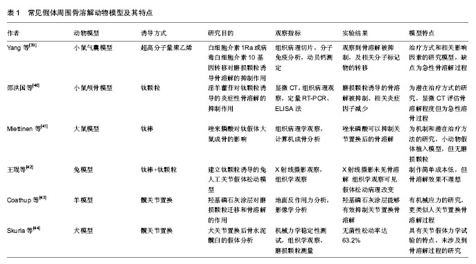
| [1] Francesca V,Matilde T,Milena F,et al.Gene expression in osteolysis: review on the identification of altered molecular pathways in preclinical and clinical studies. Int J Mol Sci. 2017;18(3):499.[2] Drynda A,Singh G,Buchhorn G,et al.Metallic wear debris may regulate CXCR4 expression in vitro and in vivo.Biomed Mater Res A. 2015;103:1940-1948.[3] Saad S,Dharmapatni A,Crotti T,et al.Semaphorin-3a, neuropilin-1 and plexin-A1 in prosthetic-particle induced bone loss Acta Biomater.2016;30:311-318.[4] Zhao YP, Wei JL, Tian QY,et al.Progranulin suppresses titanium particle induced inflammatory osteolysis by targeting TNFα signaling.Sci Rep.2016;6:20909.[5] Cordova LA,Stresing V,Gobin B,et al.Orthopaedic implant failure:Aseptic implant loosening-The contribution and future challenges of mouse models in translational research.Clin Sci. 2014;127:277-293.[6] Kurtz S,Ong K,Lau E,et al. Projections of primary and revision hip and knee arthroplasty in the United States from 2005 to 2030. Bone Joint Surg Am. 2007;89:780 -785.[7] Inacio MC,Ake CF,Paxton EW,et al.Sex and risk of hip implant failure:assessing total hip arthroplasty outcomes in the United States. JAMA Intern Med. 2013;173(6):435-441.[8] Marshall A, Ries MD, Paprosky W,et al.How prevalent are implant wear and osteolysis, and how has the scope of osteolysis changed since 2000?. J Am Acad Orthop Surg. 2008;16 (Suppl 1): S1-S6.[9] Ren W,Yang SY,Wooley PH,et al.A novel murine model of orthopaedic wear debris associated osteolysis.Scand J Rheumatol. 2004;33(5):349-357.[10] Geng D,Xu Y,Yang H,et al.Protection against titanium particle induced osteolysis by cannabinoid receptor 2 selective antagonist.Biomaterials. 2010;31(8):1996-2000.[11] 陈德胜,李燕,郭凤英,等.钛颗粒诱导小鼠植骨气囊骨溶解模型的构建[J].宁夏医科大学学报,2016,38(11):1228-1231.[12] 耿德春,徐耀增,朱雪松,等.钛颗粒诱导小鼠骨溶解模型的评价[J].中国组织工程研究, 2009,13(22):4223-4226.[13] Merkel KD,Erdmann JM,Mchugh KP,et al.Tumor necrosis factor-alpha mediates orthopedic implant osteolysis.Am J Pathol. 1999;154(2):203-210.[14] Wedemeyer C,Neuerburg C,Pfeiffer A,et al.Polyethylene particle-induced bone resorption in substance P-deficient mice.Calcif Tissue Int. 2007;80(4):268-274.[15] Shang JY,Zhan P,Jiang C,et al.Inhibitory effects of lanthanum chloride on wear particle-induced osteolysis in a mouse calvarial model.Biol Trace Elem Res. 2016;169(2):303-309.[16] Córdova LA,Trichet V,Escriou V,et al.Inhibition of osteolysis and increase of bone formation after local administration of siRNA-targeting RANK in a polyethylene particle-induced osteolysis mode.Acta Biomater.2015;13:150-158.[17] 张雨笛,严明,俞立虹,等.PI3K/Akt信号通路在磷酸三钙磨损颗粒诱导小鼠颅骨溶解中的作用[J].中国运动医学杂志, 2017,36(3): 212-217.[18] Warme B,Epstein N,Trindade M,et al.Proinflammatory mediator expression in a novel murine model of titanium particle induced intramedullary inflammation. J Biomed Mater Res B Appl Biomater. 2004;71:360-366.[19] Yang S,Yu H,Gong W,et al.Murine model of prosthesis failure for the long-term study of aseptic loosening.J Orthop Res. 2007;25:603 -611.[20] Zhang T,Yu H,Gong W,et al.The effect of osteoprotegerin gene modification on wear debris induced osteolysis in a murine model of knee prosthesis failure. Biomaterials. 2009; 30(30):6102-6108.[21] Ren W,Zhang R,Hawkins M,et al. Efficacy of periprosthetic erythromycin delivery for wear debris-induced inflammation and osteolysis.Inflamm Res. 2010;59(12):1091-1097.[22] Pap G, Machner A, Rinnert T, et al. Development and characteristics of a synovial-like interface membrane around cemented tibial hemiarthroplasties in a novel rat model of aseptic prosthesis loosening. Arthritis Rheum. 2001;44(4): 956-963.[23] Howie DW,Vernon-Roberts B,Oakeshott R, et al. A rat model of resorption of bone at the cement-bone interface in the presence of polyethylene wear particles.J Bone Joint Surg Am. 1988;7:257-263.[24] Gelb H,Schumacher H,Cuckler J,et al.In vivo inflammatory response to polymethylmethacrylate particulate debris:effect of size,morphology,and surface area.J Orthop Res.1994;12: 83-92.[25] Allen M,Brett F,Millett P,et al.The effects of particulate polyethylene at a weight-bearing bone-implant interface:a study in rats.J Bone Joint Surg Br. 1996;78: 32-37.[26] Ren W,Muzik O,Jackson N,et al.Differentiation of septic and aseptic loosening by PET with both 11C-PK11195 and 18F-FDG in rat models.Nucl Med Commun. 2012;33(7): 747-756.[27] Miettinen SS,Jaatinen J,Pelttari A,et al.Effect of locally administered zoledronic acid on injury-induced intramembranous bone regeneration and osseointegration of a titanium implant in rats.J Orthop Sci. 2009;14:431-436.[28] 丁悦,黄健斌,秦础强.人工关节无菌性松动动物模型的建立[J].实用骨科杂志,2010,16(7):510-515.[29] Im GI, Kwon BC, Lee KB. The effect of COX-2 inhibitors on periprosthetic osteolysis. Biomaterials.2004;25(2):269-275.[30] Fornasier VL, Goodman SB, Protzner K, et al. The role of implant alignment on stability and particles on periprosthetic osteolysis--A rabbit model of implant failure. J Biomed Mater Res B Appl Biomater. 2004;70(2):179-186.[31] Zhao X, Cai XZ, Shi ZL,et al,Low intensity pulsed ultrasound (LIPUS) may prevent polyethylene induced periprosthetic osteolysis in vivo.Ultrasound Med Biol. 2012;38:238-246.[32] Hu B,Cai XZ,Shi ZL,et al.Microbubble injection enhances inhibition of low-intensity pulsed ultrasound on debris-induced periprosthetic osteolysis in rabbit model.Ultrasound Med Biol.2015;41(1):177-186.[33] Lennox DW,Lewis CG,Jones LC,et al.Tissue response to motion at the bone/cement interface: a histological study in a canine model.Trans Soc Biomaterials.1987;10:133.[34] Jones LC,Frondoza C,Hungerford DS.Effect of PMMA particles and movement on an implant interface in a canine model.J Bone Joint Surg Br.2001;83(3):448-458.[35] Lind M,Overgaard S,Bunger C,et al. Improved bone anchorage of hydroxypatite coated implants compared with tricalcium-phosphate coated implants in trabecular bone in dogs.Biomaterials. 1999;20(8):803-808.[36] 严孟宁,戴尅戎,汤亭亭.假体周围骨溶解性骨缺损的转骨形态发生蛋白-2基因治疗[J].中华实验外科杂志,2008,25(6):702-704.[37] Kalia P,Coathup MJ,Oussedik S,et al.Augmentation of bone growth onto the acetabular cup surface using bonemarrow stromal cells in total hip replacement surgery. Tissue Eng Part A. 2009;15:3689-3696.[38] El-Warrak AO,Olmstead M,Apelt D,et al.An animal model for interface tissue formation in cemented hip replacements.Vet Surg. 2004;33:495-504.[39] Yang SY,Wu B,Mayton L,et al.Protective effects of IL-1Ra or vIL-10 gene transfer on a murine model of wear debris-induced osteolysis.Gene Therapy.2004;11(5):483-491.[40] 邵洪国,崔京福,朱世军,等.淫羊藿苷对钛颗粒诱导的炎症性骨溶解的抑制作用[J].中华创伤杂志,2015,31(9):850-855.[41] Miettinen SSA,Jaatinen J,Pelttari A,et al.Effect of locally administered zoledronic acid on injury-induced intramembranous bone regeneration and osseointegration of a titanium implant in rats.J Orthop Sci. 2009;14(4):431-436.[42] 王琨,王英振,夏长所,等.钛颗粒诱导的兔人工关节假体松动模型制备[J].青岛大学医学院学报,2013,49(5):399-402.[43] Coathup MJ,Blackburn J,Goodship AE,et al.Role of hydroxyapatite coating in resisting wear particle migration and osteolysis around acetabular components.Biomaterials. 2005; 26(26):4161-4169.[44] Skurla CP,James SP.Assessing the dog as a model for human total hip replacement:analysis of 38 postmortem- retrieved canine cemented acetabular components.J Bone Joint Surg. 2005;87(1):120-127.[45] Ren W,Markel DC,Schwendener R,et al. Macrophage depletion diminishes implant-wear-induced inflammatory osteolysis in a mouse model. J Biomed Mater Res A. 2008; 85(4):1043-1051.[46] Zhang HW,Peng L,Li WB,et al.The role of RANKL/RANK/OPG system in the canine model of hip periprosthetic infection osteolysis. Int J Artif Organs. 2016; 39(12): 619-624.[47] Ma T,Ortiz S,Huang Z,et al.In vivo murine model of continuous intramedullary infusion of particles-A preliminary study.Biomed Mater Res B Appl Biomater. 2009;88(1): 250-253.[48] 刘铭,张健,魏小兰,等.改良型磨损颗粒诱导小鼠颅骨溶解模型的构建[J].中国老年学,2013,33(23):5927-5930.[49] 万睿,李平,周园东,等.纱布纤维可否在小鼠气囊植骨模型中诱发炎性骨溶解的研究[J].中国全科医学,2013,16(7):648-652.[50] Mcgonigle P,Ruggeri B.Animal models of human disease: Challenges in enabling translation. Biochem Pharmacol. 2014; 87(1):162-171. |
.jpg) 文题释义:
文题释义:
.jpg)
.jpg) 文题释义:
文题释义: