中国组织工程研究 ›› 2018, Vol. 22 ›› Issue (6): 921-926.doi: 10.3969/j.issn.2095-4344.0069

• 生物材料基础实验 basic experiments of biomaterials • 上一篇    下一篇

富血小板血浆与肌腱干细胞共混物干预跟腱病模型兔肌腱组织形态及金属蛋白酶1的表达

咸  杰,何本祥,吴  骁,檀亚军
  

  1. 成都体育学院,四川省成都市  610041
  • 收稿日期:2017-09-08 出版日期:2018-02-28 发布日期:2018-02-28
  • 通讯作者: 何本祥,博士,主任医师,博士生导师,成都体育学院,四川省成都市 610041
  • 作者简介:咸杰,男,1989年生,汉族,硕士,主要从事运动创伤防治研究。
  • 基金资助:
    四川省科技厅重点项目(2016JY0052);四川省科技厅支持项目(2014SZ0003);成都体育学院硕士点建设基金项目(16SSZX01)

Autologous platelet-rich plasma in combination with tendon stem cells to treat tendinopathy in a rabbit model: histomorphological changes of the tendon tissue and matrix metalloproteinase 1 expression

Xian Jie, He Ben-xiang, Wu Xiao, Tan Ya-jun
  

  1. Chengdu Sport Institute, Chengdu 610041, Sichuan Province, China
  • Received:2017-09-08 Online:2018-02-28 Published:2018-02-28
  • Contact: He Ben-xiang, M.D., Chief physician, Doctoral supervisor, Chengdu Sport Institute, Chengdu 610041, Sichuan Province, China
  • About author:Xian Jie, Master, Chengdu Sport Institute, Chengdu 610041, Sichuan Province, China
  • Supported by:
    the Key Project of Science and Technology Department of Sichuan Province, No. 2016JY0052; Science and Technology Support Project of Science and Technology Department of Sichuan Province, No. 2014SZ0003; the Master Project of Chengdu Sport Institute, No. 16SSZX01

摘要:

文章快速阅读:


文题释义:
富血小板血浆:是自体血小板的浓缩后的产物,通过采集外周血后二次离心后制备,其中的α-颗粒可释放许多生长因子和细胞因子,其生长因子含量高于正常血浆的3-5 倍,主要的生长因子包括有转化生长因子β、胰岛素样生长因子1、血管内皮生长因子等,这些因子能够在组织修复工程中发挥关键作用。
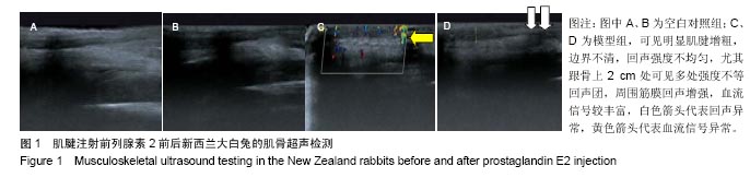
前列腺素E2制作跟腱病模型的机制:可能是前列腺素E2能够使肌腱组织中Ⅰ、Ⅲ型胶原细胞外基质发生改变,使Ⅰ型胶原纤维含量下降的同时,Ⅲ型胶原表达上升,降低其生物力学特性,其表现与因退变所致的改变一致;同时前列腺素E2能够刺激炎性递质的产生,增加血管通透性,促进炎性细胞的趋化与浸润,这一过程与肌腱病初期炎性改变吻合。
 
背景:富血小板血浆针对组织重建修复的研究及应用一直是医学及生物工程学的研究热点。
目的:观察富血小板血浆与肌腱干细胞共混物对肌腱病兔肌腱组织形态及金属蛋白酶1表达的影响。
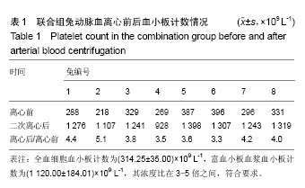
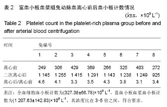
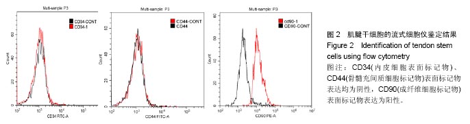
方法:将40只新西兰大白兔随机分为2组,模型组(n=32)在距左跟骨附着点约2 cm处注射前列腺素E2,1次/周,共4周,建立肌腱病动物模型;空白对照组(n=8)相同部位注射等量的生理盐水。4周后,将模型组随机分为4组,联合组于跟腱局部注射自体富血小板血浆与自体肌腱干细胞混合物,干细胞组注射自体肌腱干细胞,富血小板血浆组注射自体富血小板血浆,模型对照组注射等量的生理盐水,每3周1次,注射2次。注射6周后取跟腱组织标本,进行组织学检查及金属蛋白酶1检测。
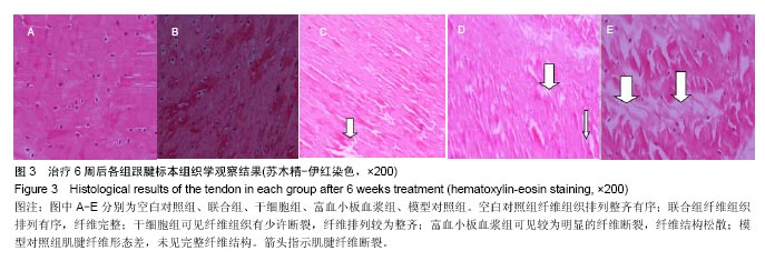
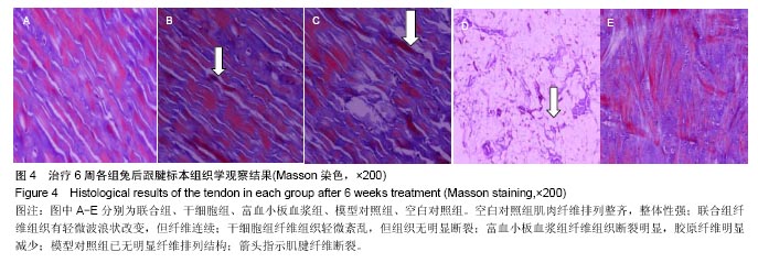
结果与结论:①苏木精-伊红染色:联合组纤维组织排列有序,纤维完整;干细胞组纤维组织有少许断裂,纤维排列较为整齐;富血小板血浆组纤维断裂明显,纤维结构松散;模型对照组肌腱纤维形态差,未见完整纤维结构,炎性细胞浸润明显;②Masson染色:联合组纤维组织有轻微波浪状改变,但纤维连续;干细胞组纤维组织轻微紊乱,组织无明显断裂,炎性细胞浸润明显;富血小板血浆组纤维组织断裂明显,胶原纤维明显减少,炎性细胞浸润明显;模型对照组已无明显纤维排列结构,炎性细胞浸润明显;③金属蛋白酶1水平:干细胞组、富血小板血浆组、模型对照组金属蛋白酶1水平高于空白对照组(P < 0.05),联合组金属蛋白酶1水平与空白对照组接近(P > 0.05);④结果表明:富血小板血浆与肌腱干细胞共混物可通过抑制肌腱细胞基质及胶原降解的恶性循环、下调肌腱细胞中金属蛋白酶1表达,延缓肌腱病炎性反应及退行性改变。

关键词: 富血小板血浆, 肌腱干细胞, 肌腱病, 运动医学, 金属蛋白酶, 动物模型, 组织修复, 生物材料

Abstract:

BACKGROUND: The research and application of platelet-rich plasma in tissue regeneration and restoration have always been an issue of concern in the medicine and bioengineering fields.
OBJECTIVE: To analyze the effects of platelet-rich plasma in combination with tendon stem cells on histomorphology change and matrix metalloproteinase 1 expression of the tendon tissues in a rabbit model of Achilles tendinopathy.
METHODS: Forty New Zealand white rabbits were randomly divided into model group (n=32) and blank control group (n=8). In the model group, the animals were injected about 2 cm distant to the attachment point of the left calcaneus with prostaglandin E2 (once a week, for totally 4 weeks) to make the animal model of tendinopathy. The rabbits in the blank control group were injected the equal amount of normal saline. After 4 weeks, model rabbits were randomly divided into four subgroups: combination group, tendon stem cell group, platelet-rich plasma group and model control group, with eight rabbits in each group. Platelet-rich plasma and tendon stem cells, alone or in combination, and normal saline were injected into the corresponding group, twice with an interval of 3 weeks. At 6 weeks after injection, the tendon tissue was collected and stained for histological examination and detection of matrix metalloproteinase 1 expression.
RESULTS AND CONCLUSION: (1) Hematoxylin-eosin staining: the tendon fibers in the combinationgroup were intact and arranged orderly; in the tendon stem cell group, the tendon fibers were almost arranged orderly despite some fractured fibers; in the platelet-rich plasma group, fiber breakage and loose fiber structure were observed; in the model control group, there were no intact tendon fibers, with the presence of inflammatory cell filtration. (2) Masson staining: The tendon fibers in the combination group had slight wave-shaped changes but the fibers were not cut off; in the tendon stem cell group, the tendon fibers were slightly in disorder, but with the intact structure, and obvious inflammatory cell filtration was observed; in the platelet-rich plasma group, fiber breakage, reduced collagen fibers and inflammatory cell filtration were obviously observed; in the model control group, there were no intact tendon fibers, and inflammatory cell filtration was clearly visible. (3) The expression of matrix metalloproteinase 1: Compared with the blank control group, the expression of matrix metalloproteinase 1 was significantly higher in the other groups except the combination group (P < 0.05). There was no significant difference in the expression of matrix metalloproteinase 1 between the combination group and blank control group (P > 0.05). To conclude, the combination of platelet-rich plasma and tendon stem cells can inhibit the vicious cycle of degeneration of collagen and extracellular matrix, reduce the expression of matrix metalloproteinase 1 in tenocytes, and delay inflammation responses and degeneration due to tendinopathy.

Key words: Platelet-Rich Plasma, Stem Cells, Tendinopathy, Metalloproteases, Tissue Engineering

中图分类号: