[1] 姜宗来.发展生物力学 造福人类健康——“十四五”我国生物力学研究发展战略思考[J].医用生物力学,2021,36(5):671-675.
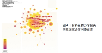
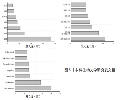
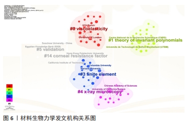
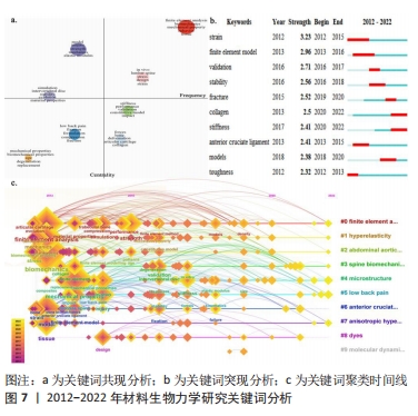
[2] 陈振银,吕永钢. 材料生物力学2021年研究进展[J].医用生物力学, 2022,37(2):211-218.
[3] 于振涛,余森,程军,等.新型医用钛合金材料的研发和应用现状[J].金属学报,2017,53(10):1238-1264.
[4] 魏俊超,李晓娜.圆锥角膜生物力学研究进展[J].太原理工大学学报, 2022,53(3):443-449.
[5] 康巍,徐鹏,卜伟平,等.生物软组织力学测试及相关理论研究[J].兵工学报,2022,43(9):2164-2171.
[6] 林伟坚,张博文,汪卫华.从全球气候变化、制造业产业升级、国家安全及材料基因工程维度探讨材料科学发展趋势[J].中国科学院院刊, 2022,37(3):336-342.
[7] 万遂人,顾晓松,骆清铭,等.生物医学工程发展方向和我国高端医疗器械突破点[J] 广西医科大学学报, 2023,40(4):543-548.
[8] SIONKOWSKA A. Collagen Based Materials for Biomedical Applications: Preparation and Properties. Mater Sci Forum. 2012;706-709(1):595-599.
[9] RACHEL T, ZEIKE T. Photoelastic materials and methods for tissue biomechanics applications. Opti Eng. 2015;54(8):081208.
[10] YANG HL, XU H, WU YK, et al. Methods of Porous Biomedical Material Fabrication. Adv Mater Res. 2013;750-752(3):1468-1471.
[11] 贾巍,张满栋,陈维毅,等.股骨假体材料对人工膝关节置换性能的影响[J].中国组织工程研究,2021,25(10):1477-1481.
[12] 黄茂茂,胡月,王彬川,等.缺血性脑卒中康复近10年国际文献计量学及可视化分析[J].中国组织工程研究,2021,25(23):3725-3733.
[13] DREISCHARF M, ZANDER T, SHIRAZI-ADL A, et al. Comparison of eight published static finite element models of the intact lumbar spine: Predictive power of models improves when combined together. J Biomech. 2014;47(8):1757-1766.
[14] SON K, GUASTO JS, STOCKER R. Guasto,Roman Stocker. Bacteria can exploit a flagellar buckling instability to change direction. Nat Phys. 2013;9(8):494-498.
[15] 李文斌,洪靖.近十年国内外体育传播研究述评[J].情报探索,2023, 305(3):128-134.
[16] 赵稼祥,吴方才.国外生物材料和加工的发展[J].宇航材料工艺,1992, 21(1):48-49.
[17] HASAN I, KEILIG L, BOURAUEL C, et al. The effect of screw preload and framework material on the success of cementable fixed partial prostheses: A finite element study. Ann Anat. 2015;199:58-66.
[18] YANG M, XIANG D, WANG S, et al. In Vitro Studies for Investigating Creep of Intervertebral Discs under Axial Compression: A Review of Testing Environment and Results. Materials (Basel). 2022;15(7):2500.
[19] 钱蕾,欧阳钧.两种新型椎弓根螺钉固定强度的体外生物力学研究[J].医用生物力学,2021,36(S1):22.
[20] TANG M, TIWARI SK, AGRAWAL K, et al. Rapid 3D Bioprinting of Glioblastoma Model Mimicking Native Biophysical Heterogeneity. Small. 2021;17(15): 2006050.
[21] BAY BK, SMITH TS, FYHRIE DP, et al. Digital volume correlation: Three-dimensional strain mapping using X-ray tomography. Exp Mech. 1999;39(3): 217-226.
[22] 丁学东.文献计量学基础[M].北京: 北京大学出版社,1992:204-209.
[23] MA Z, VYHLIDAL MJ, LI DX, et al. Mechano-bioengineering of the knee meniscus. Am J Physiol Cell Physiol. 2022;323(6):C1652-C1663.
[24] LI RL, RUSS J, PASCHALIDES C, et al. Mechanical considerations for polymeric heart valve development: Biomechanics, materials, design and manufacturing. Biomaterials. 2019;225:119493.
[25] PEISKER H, MICHELS J, GORB SN. Evidence for a material gradient in the adhesive tarsal setae of the ladybird beetle Coccinella septempunctata. Nat Commun. 2013;4:1661.
[26] 刘镕阁,徐雁.三维有限元建模分析在髋关节撞击综合征诊疗中的应用研究进展[J].中国运动医学杂志,2021,40(4):317-321.
[27] 周玉,龙小安,李宁,等.有限元法分析髌腱炎状态时的生物力学变化[J].中国组织工程研究,2020,24(8):1280-1286.
[28] KUMAR A, SHITOLE P, GHOSH R, et al. Experimental and numerical comparisons between finite element method, element-free Galerkin method, and extended finite element method predicted stress intensity factor and energy release rate of cortical bone considering anisotropic bone modelling. Proc Inst Mech Eng H. 2019;233(8):823-838.
[29] FUKSA AK, RACHOWICZ W. Numerical simulations of arteries with an adaptive finite element method. J Theor App Mech-Pol. 2014;52(4):917-925.
[30] WAN C, HAO Z, WEN S. The Effect of the Variation in ACL Constitutive Model on Joint Kinematics and Biomechanics Under Different Loads: A Finite Element Study. J Biomech Eng. 2013;135(4):041002.
[31] CHOKHANDRE S, HALLORAN JP, VAN DEN BOGERT AJ, et al. A Three-Dimensional Inverse Finite Element Analysis of the Heel Pad. J Biomech Eng. 2012;134(3):031002.
[32] GUO LX, LI WJ. Finite element modeling and static/dynamic validation of thoracolumbar-pelvic segment. Comput Methods Biomech Biomed Engin. 2020;23(2):69-80.
[33] LIU J, QU X, LIU Y. Influence of Load Knowledge on Biomechanics of Asymmetric Lifting. Int J Environ Res Public Health. 2022;19(6):3207.
[34] WAN C, HAO ZX, WEN SZ. The effect of the material property change of anterior cruciate ligament by ageing on joint kinematics and biomechanics under tibial varus/valgus torques. Biomed Mater Eng. 2014;24(1): 1375-1382.
[35] HOU JY, ZHANG SQ, KE L. The Application of Metal Materials in Exercise-Induced Bone Injury. Adv Mater Res. 2013;675(3):205-208.
[36] LU C,FAN Y,YU G, et al. Asymptomatic foot and ankle structural injuries: a 3D imaging and finite element analysis of elite fencers. BMC Sports Sci Med Rehabil. 2022;14(1):50.
[37] SEYED SAS, FARZAN G, IMAN ZO. A scaled boundary finite element formulation for solving plane-strain viscoelastic problems. Eur J Mech A Solids. 2022;96(8):104755.
[38] CORTEZ S, FREITAS FL, COMPLETO A, et al. A 3D finite element model to predict the arcade-like collagen structure in a layered PCL scaffold for cartilage tissue engineering. Comput Methods Biomech Biomed Engin. 2017;20(sup1):47-48.
[39] MELONI GR, FISHER MB, STOECKL BD, et al. Biphasic Finite Element Modeling Reconciles Mechanical Properties of Tissue-Engineered Cartilage Constructs Across Testing Platforms. Tissue Eng Part A. 2017;23(13-14): 663-674.
[40] BAS O, DE-JUAN-PARDO EM, MEINERT C, et al. Biofabricated soft network composites for cartilage tissue engineering. Biofabrication. 2017;9(2): 025014.
[41] WEISS JA, GARDINER JC. Computational modeling of ligament mechanics. Crit Rev Biomed Eng. 2001;29(3):303-371.
[42] SPARKS JL, VAVALLE NA, KASTING KE, et al. Use of silicone materials to simulate tissue biomechanics as related to deep tissue injury. Adv Skin Wound Care. 2015;28(2):59-68.
[43] GRYKO A, PROCHOR P, SAJEWICZ E. Finite element analysis of the influence of porosity and pore geometry on mechanical properties of orthopaedic scaffolds. J Mech Behav Biomed Mater. 2022;132:105275.
[44] 杨顺清,毛萱.无机生物材料学[M].广州:华南理工大学出版社,2008: 3-15.
[45] CIAPETTI G, DI POMPO G, AVNET S, et al. Osteoclast differentiation from human blood precursors on biomimetic calcium-phosphate substrates. Acta Biomater. 2017;50:102-113.
[46] RODRIGUEZ-FONTAN F. Fracture healing, the diamond concept under the scope: hydroxyapatite and the hexagon. Medicina. 2022;82(5): 764-769.
[47] DOMENICI P, SEEBACHER F. The impacts of climate change on the biomechanics of animals: Themed Issue Article: Biomechanics and Climate Change. Conserv Physiol. 2020;8(1):coz102.
[48] ESTER C, FACUNDO JB, SERGIO O. A generalized finite-strain damage model for quasi-incompressible hyperelasticity using hybrid formulation. Int J Numer Methods Eng. 2015;105(10):781-800.
[49] SUYA PREM ANAND P, ARUNACHALAM N, VIJAYARAGHAVAN L. Investigation on Grindability of Medical Implant Material Using a Silicon Carbide Wheel with Different Cooling Conditions. Procedia Manufacturing. 2017;10(9): 417-428.
[50] DISCHER D, DONG C, FREDBERG JJ, et al. Biomechanics: Cell Research and Applications for the Next Decade. Ann Biomed Eng. 2009;37(5):847-859.
[51] RENNEKAMP B, KUTZKI F, OBARSKA-KOSINSKA A, et al. Hybrid Kinetic Monte Carlo/Molecular Dynamics Simulations of Bond Scissions in Proteins. J Chem Theory Comput. 2020;16(1):553-563.
[52] LAI ZB, BAI R, LEI Z, et al. Interfacial mechanical behaviour of protein–mineral nanocomposites: A molecular dynamics investigation. J Biomech. 2018;73:161-167.
[53] JIANG LG, WU HA, ZHOU XZ, et al. Coarse-Grained Molecular Dynamics Simulation of a Red Blood Cell. Chin Phys Lett. 2010;27(2):028704.
[54] FILIPOWSKA J, TOMASZEWSKI KA, NIEDŹWIEDZKI Ł, et al. The role of vasculature in bone development, regeneration and proper systemic functioning. Angiogenesis. 2017;20(3):291-302.
[55] BENJAMIN SH, SHINJI I, DANIEL JR, et al. Continuum mechanical parameterisation of cytoplasmic dynein from atomistic simulation. Methods. 2021;185(2):39-48.
[56] MICHAEL ES, RICHARD AR,VIRGINIA LF. A poroelastic finite element model of the bone–cartilage unit to determine the effects of changes in permeability with osteoarthritis. Comput Methods Biomech Biomed Engin. 2017;20(3):319-331. |