[1] NOURISSAT G, BERENBAUM F, DUPREZ D. Tendon injury: from biology to tendon repair. Nat Rev Rheumatol. 2015;11(4):223-233.
[2] TEMPLEHOF S, RUPP S, SEIL R. Age-related prevalence of rotator cuff tears in asymptomatic shoulders. J Shoulder Elbow Surg. 1999;8(4):296-299.
[3] LUI PP. Stem cell technology for tendon regeneration: current status, challenges, and future research directions. Stem Cells Cloning. 2015;8: 163-174.
[4] LIM WL, LIAU LL, NG MH, et al. Current progress in tendon and ligament tissue engineering. Tissue Eng Regen Med. 2019;16(6):549-571.
[5] FU SC, ROLF C, CHEUK YC, et al. Deciphering the pathogenesis of
tendinopathy: a three-stages process. Sports Med Arthrosc Rehabil Ther Technol. 2010;2:30.
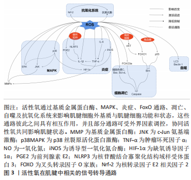
[6] 李凯群.高胆固酵通过激活活性氧介导的NF-кB通路抑制肌腱干细胞的腱系分化[D].广州:南方医科大学,2019.
[7] ABATE M, DI CARLO L, COCCO G, et al. Oxidative stress and abnormal tendon sonographic features in elite soccer players (a pilot study). Rev Bras Ortop. 2021;56(4):432-437.
[8] NOH KC, PARK SH, YANG CJ, et al. Involvement of synovial matrix degradation and angiogenesis in oxidative stress-exposed degenerative rotator cuff tears with osteoarthritis. J Shoulder Elbow Surg. 2018;27(1):141-150.
[9] YOSHIDA K, ITOIGAWA Y, WADA T, et al. Association of superoxide-induced oxidative stress with rotator cuff tears in juman patients. J Orthop Res. 2020;38(1):212-218.
[10] ITOIGAWA Y, YOSHIDA K, NOJIRI H, et al. Association of recurrent tear after arthroscopic rotator cuff repair and Superoxide-Induced oxidative stress. Am J Sports Med. 2021;49(8):2048-2055.
[11] FU SC, YEUNG MY, ROLF CG, et al. Hydrogen peroxide induced tendinopathic changes in a rat model of patellar tendon injury. J Orthop Res. 2018;36(12): 3268-3274.
[12] RAY PD, HUANG BW, TSUJI Y. Reactive oxygen species (ROS) homeostasis and redox regulation in cellular signaling. Cell Signal. 2012;24(5):981-990.
[13] GALLORINI M, BERARDI AC, GISSI C, et al. Nrf2-mediated cytoprotective effect of four different hyaluronic acids by molecular weight in human tenocytes. J Drug Target. 2020;28(2):212-224.
[14] LIU YC, WANG HL, HUANG YZ, et al. Alda-1, an activator of ALDH2, ameliorates Achilles tendinopathy in cellular and mouse models. Biochem Pharmacol. 2020;175:113919.
[15] BESTWICK CS, MAFFULLI N. Reactive oxygen species and tendinopathy: do they matter? Br J Sports Med. 2004;38(6):672-674.
[16] LUI PPY, ZHANG X, YAO S, et al. Roles of oxidative stress in acute tendon injury and degenerative tendinopathy-a target for intervention. Int J Mol Sci. 2022;23(7):3571.
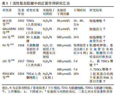
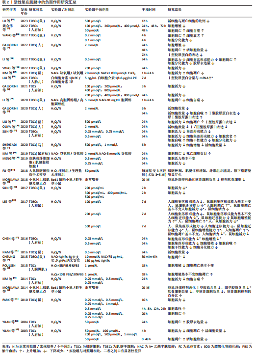
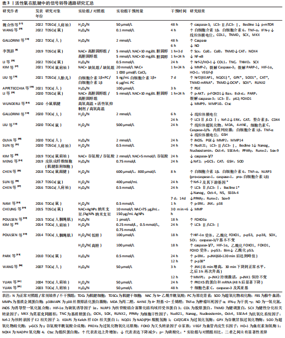
[17] 魏合伟,郑维蓬,刘治军,等.化应激诱导肩袖肌腱干细胞自噬的表达[J].中国组织工程研究,2022,26(31):4954-4961.
[18] GALLORINI M, BERARDI AC, RICCI A, et al. Dual acting carbon monoxide releasing molecules and carbonic anhydrase inhibitors differentially modulate inflammation in human tenocytes. Biomedicines. 2021;9(2):141.
[19] LEE YW, FU SC, YEUNG MY, et al. Effects of redox modulation on cell proliferation, viability, and migration in cultured rat and human tendon progenitor cells. Oxid Med Cell Longev. 2017;2017:8785042.
[20] POULSEN RC, KNOWLES HJ, CARR AJ, et al. Cell differentiation versus cell death: Extracellular glucose is a key determinant of cell fate following oxidative stress exposure. Cell Death Dis. 2014;5(2):e1074.
[21] ZHANG C, ZHANG E, YANG L, et al. Histone deacetylase inhibitor treated cell sheet from mouse tendon stem/progenitor cells promotes tendon repair. Biomaterials. 2018;172:66-82.
[22] LU K, ZHOU M, WANG L, et al. N-Acetyl-L-cysteine facilitates tendon repair and promotes the tenogenic differentiation of tendon stem/progenitor cells by enhancing the integrin α5/β1/PI3K/AKT signaling. BMC Mol Cell Biol. 2023;24(1):1.
[23] WANG S, YAO Z, ZHANG X, et al. Energy-supporting enzyme-mimic
nanoscaffold facilitates tendon regeneration based on a mitochondrial protection and microenvironment remodeling strategy. Adv Sci (Weinh). 2022;9(31):e2202542.
[24] GALLORINI M, ANTONETTI LAMORGESE PASSERI C, CATALDI A, et al. Hyaluronic acid alleviates oxidative stress and apoptosis in human tenocytes via Caspase 3 and 7. Int J Mol Sci. 2022;23(15):8817.
[25] LI X, SU Z, SHEN K, et al. Eugenol-Preconditioned mesenchymal stem Cell-Derived extracellular vesicles promote antioxidant capacity of tendon stem cells in vitro and in vivo. Oxid Med Cell Longev. 2022;2022:3945195.
[26] SONG K, JIANG T, PAN P, et al. Exosomes from tendon derived stem cells promote tendon repair through miR-144-3p-regulated tenocyte proliferation and migration. Stem Cell Res Ther. 2022;13(1):80.
[27] KIM RJ, AN SH, GWARK JY, et al. Antioxidant effects on hypoxia-induced oxidative stress and apoptosis in rat rotator cuff fibroblasts. Eur Cell Mater. 2021;41:680-693.
[28] LIU R, ZHOU B, ZHANG H, et al. Inhibition of ROS activity by controlled release of proanthocyanidins from mesoporous silica nanocomposites effectively ameliorates heterotopic ossification in tendon. Chem Eng J. 2021;420(1):129415.
[29] LI K, DENG Y, DENG G, et al. High cholesterol induces apoptosis and autophagy through the ROS-activated AKT/FOXO1 pathway in tendon-derived stem cells. Stem Cell Res Ther. 2020;11(1):131.
[30] OLIVA F, GALLORINI M, PASSERI CAL, et al. Conjugation with methylsulfonylmethane improves hyaluronic acid anti-inflammatory activity in a hydrogen peroxide-exposed tenocyte culture in vitro model. Int J Mol Sci. 2020;21(21):1-15.
[31] SUN Y, CHEN H, YE H, et al. Nudt21-mediated alternative polyadenylation of HMGA2 3’-UTR impairs stemness of human tendon stem cell. Aging (Albany NY). 2020;12(18):18436-18452.
[32] SHEHZADI S, JAVED M, AWAN SJ, et al. Developing a novel, efficient and cost effective tissue injury model (in-vitro) on equine tendon fibroblasts. Pak Vet J. 2020;40(3):360-364.
[33] KIM RJ, HAH YS, GWARK JY, et al. N-acetylcysteine reduces glutamate-induced cytotoxicity to fibroblasts of rat supraspinatus tendons. Connect Tissue Res. 2019;60(5):431-443.
[34] MENG J, YU P, TONG J, et al. Hydrogen treatment reduces tendon adhesion and inflammatory response. J Cell Biochem. 2019;120(2):1610-1619.
[35] MORIKAWA D, NOJIRI H, ITOIGAWA Y, et al. Antioxidant treatment with vitamin C attenuated rotator cuff degeneration caused by oxidative stress in Sod1-deficient mice. JSES Open Access. 2018;2(1):91-96.
[36] SUN W, MENG J, WANG Z, et al. Proanthocyanidins attenuation of H2O2-Induced oxidative damage in Tendon-Derived stem cells via upregulating nrf-2 signaling pathway. Biomed Res Int. 2017;2017:7529104.
[37] CHEN H, GE HA, WU GB, et al. Autophagy prevents oxidative Stress-Induced loss of Self-Renewal capacity and stemness in human tendon stem cells by reducing ROS accumulation. Cell Physiol Biochem. 2016;39(6):2227-2238.
[38] NAM DC, HAH YS, NAM JB, et al. Cytoprotective mechanism of cyanidin and delphinidin against oxidative Stress-Induced tenofibroblast death. Biomol Ther. 2016;24(4):426-432.
[39] CHEUNG TS, LAU PM, LU H, et al. Cytotoxic and sublethal effects of silver nanoparticles on tendon-derived stem cells - implications for tendon engineering. Toxicol Res. 2015;5(1):318-330.
[40] POULSEN RC, CARR AJ, HULLEY PA. Cell proliferation is a key determinant of the outcome of FOXO3a activation. Biochem Biophys Res Commun. 2015;462(1):78-84.
[41] KIM RJ, HAH YS, SUNG CM, et al. Do antioxidants inhibit oxidative-stress-induced autophagy of tenofibroblasts? J Orthop Res. 2014;32(7):937-943.
[42] MORIKAWA D, ITOIGAWA Y, NOJIRI H, et al. Contribution of oxidative stress to the degeneration of rotator cuff entheses. J Shoulder Elbow Surg. 2014;23(5):628-635.
[43] PARK HB, HAH YS, YANG JW, et al. Antiapoptotic effects of anthocyanins on rotator cuff tenofibroblasts. J Orthop Res. 2010;28(9):1162-1169.
[44] YUAN J, MURRELL GA, TRICKETT A, et al. Overexpression of antioxidant enzyme peroxiredoxin 5 protects human tendon cells against apoptosis and loss of cellular function during oxidative stress. Biochim Biophys Acta. 2004;1693(1):37-45.
[45] YUAN J, MURRELL GA, TRICKETT A, et al. Involvement of cytochrome c release and caspase-3 activation in the oxidative stress-induced apoptosis in human tendon fibroblasts. Biochim Biophys Acta. 2003;1641(1):35-41.
[46] JAMES S, SCHUIJERS J, DAFFY J, et al. Ciprofloxacin reduces tenocyte viability and proteoglycan synthesis in short-term explant cultures of equine tendon. PeerJ. 2021;9:e12003.
[47] PIZZINO G, IRRERA N, CUCINOTTA M, et al. Oxidative stress: harms and benefits for human health. Oxid Med Cell Longev. 2017;2017:8416763.
[48] CHEN J, WANG A, XU J, et al. In chronic lateral epicondylitis, apoptosis and autophagic cell death occur in the extensor carpi radialis brevis tendon. J Shoulder Elbow Surg. 2010;19(3):355-362.
[49] SCREEN HRC, BERK DE, KADLER KE, et al. Tendon functional extracellular matrix. Journal of Orthopaedic Research. 2015;33(6):793-799.
[50] BONNANS C, CHOU J, WERB Z. Remodelling the extracellular matrix in development and disease. Nat Rev Mol Cell Biol. 2014;15(12):786-801.
[51] ROZARIO T, DESIMONE DW. The extracellular matrix in development and morphogenesis: a dynamic view. Dev Biol. 2010;341(1):126-140.
[52] BHABRA G, WANG A, EBERT JR, et al. Lateral elbow tendinopathy: development of a pathophysiology-based treatment algorithm. Orthop J Sports Med. 2016;4(11):2325967116670635.
[53] APPETECCHIA F, CONSALVI S, BERRINO E, et al. A Novel class of dual-acting DCH-CORMs counteracts oxidative stress-induced inflammation in human primary tenocytes. Antioxidants (Basel). 2021;10(11):1828.
[54] WUNDERLI SL, BLACHE U, BERETTA PICCOLI A, et al. Tendon response to matrix unloading is determined by the patho-physiological niche. Matrix Biol. 2020;89:11-26.
[55] CHEN Q, ZHOU J, ZHANG B, et al. Cyclic stretching exacerbates tendinitis by enhancing NLRP3 inflammasome activity via F-actin depolymerization. Inflammation. 2018;41(5):1731-1743.
[56] WANG F, MURRELL GA, WANG MX. Oxidative stress-induced c-Jun N-terminal kinase (JNK) activation in tendon cells upregulates MMP1 mRNA and protein expression. J Orthop Res. 2007;25(3):378-389.
[57] DEL BUONO A, OLIVA F, OSTI L, et al. Metalloproteases and tendinopathy. Muscles Ligaments Tendons J. 2013;3(1):51-57.
[58] DONG SQ, XU HZ, XIA XB, et al. Activation of the ERK 1/2 and STAT3 signaling pathways is required for 661W cell survival following oxidant injury. Int J Ophthalmol. 2012;5(2):138-142.
[59] PAXTON JZ, HAGERTY P, ANDRICK JJ, et al. Optimizing an intermittent stretch paradigm using ERK1/2 phosphorylation results in increased collagen synthesis in engineered ligaments. Tissue Eng Part A. 2012;18(3-4):277-284.
[60] JOHNSON G L, LAPADAT R. Mitogen-activated protein kinase pathways mediated by ERK, JNK, and p38 protein kinases. Science. 2002;298(5600): 1911-1912.
[61] POULSEN RC, CARR AJ, HULLEY PA. Protection against glucocorticoid-induced damage in human tenocytes by modulation of ERK, Akt, and forkhead signaling. Endocrinology. 2011;152(2):503-514.
[62] MARTIN P, POGNONEC P. ERK and cell death: cadmium toxicity, sustained ERK activation and cell death. FEBS J. 2010;277(1):39-46.
[63] SUGIURA R, SATOH R, TAKASAKI T. ERK: A double-edged sword in cancer. ERK-dependent apoptosis as a potential therapeutic strategy for cancer. Cells. 2021;10(10):2509.
[64] VENTURA JJ, HÜBNER A, ZHANG C, et al. Chemical genetic analysis of the time course of signal transduction by JNK. Mol Cell. 2006;21(5):701-710.
[65] JIAO X, ZHANG Y, LI W, et al. HIF-1αinhibition attenuates severity of Achilles tendinopathy by blocking NF-κB and MAPK pathways. Int Immunopharmacol. 2022;106:108543.
[66] KIM EK, CHOI EJ. Pathological roles of MAPK signaling pathways in human diseases. Biochim Biophys Acta. 2010;1802(4):396-405.
[67] KESAVARDHANA S, MALIREDDI RKS, KANNEGANTI TD. Caspases in cell death, inflammation, and gasdermin-induced pyroptosis. Ann Rev Immunol. 2020;38:567-595.
[68] GREEN DR. Apoptotic pathways: the roads to ruin. Cell. 1998;94(6):695-698.
[69] YUAN J, WANG MX, MURRELL GA. Cell death and tendinopathy. Clin Sports Med. 2003;22(4):693-701.
[70] KUDRYAVTSEVA AV, KRASNOV GS, DMITRIEV AA, et al. Mitochondrial dysfunction and oxidative stress in aging and cancer. Oncotarget. 2016; 7(29):44879-44905.
[71] XIAO M, ZHONG H, XIA L, et al. Pathophysiology of mitochondrial lipid oxidation: role of 4-hydroxynonenal (4-HNE) and other bioactive lipids in mitochondria. Free Radic Biol Med. 2017;111:316-327.
[72] XING YQ, LI A, YANG Y, et al. The regulation of FOXO1 and its role in disease progression. Life Sci. 2018;193:124-131.
[73] WU B, CHEN J, DELA ROSA T, et al. Cellular response and extracellular matrix breakdown in rotator cuff tendon rupture. Arch Orthop Trauma Surg. 2011;131(3):405-411.
[74] BIASIZZO M, KOPITAR-JERALA N. Interplay between NLRP3 inflammasome and autophagy. Front Immunol. 2020;11:591803.
[75] LOIACONO C, PALERMI S, MASSA B, et al. Tendinopathy: pathophysiology, therapeutic options, and role of nutraceutics. a narrative literature review. Medicina (Kaunas). 2019;55(8):447.
[76] CILLI F, KHAN M, FU F, et al. Prostaglandin E2 affects proliferation and collagen synthesis by human patellar tendon fibroblasts. Clin J Sport Med. 2004;14(4):232-236.
[77] ZHANG J, WANG JH. Production of PGE(2) increases in tendons subjected to repetitive mechanical loading and induces differentiation of tendon stem cells into non-tenocytes. J Orthop Res. 2010;28(2):198-203.
[78] MILLAR NL, MURRELL GA, MCINNES IB. Inflammatory mechanisms in tendinopathy - towards translation. Nat Rev Rheumatol. 2017;13(2):110-122.
[79] SCHULZE-TANZIL G, AL-SADI O, WIEGAND E, et al. The role of pro-inflammatory and immunoregulatory cytokines in tendon healing and rupture: new insights. Scand J Med Sci Sports. 2011;21(3):337-351.
[80] JOHN T, LODKA D, KOHL B, et al. Effect of pro-inflammatory and immunoregulatory cytokines on human tenocytes. J Orthop Res. 2010; 28(8):1071-1077.
[81] BACKMAN LJ, ERIKSSON DE, DANIELSON P. Substance P reduces TNF-α-induced apoptosis in human tenocytes through NK-1 receptor stimulation. Br J Sports Med. 2014;48(19):1414-1420.
[82] ACKERMANN PW, DOMEIJ-ARVERUD E, LECLERC P, et al. Anti-inflammatory cytokine profile in early human tendon repair. Knee Surg Sports Traumatol Arthrosc. 2013;21(8):1801-1806.
[83] LIN TW, CARDENAS L, GLASER DL, et al. Tendon healing in interleukin-4 and interleukin-6 knockout mice. J Biomech. 2006;39(1):61-69.
[84] PALMER RM, ASHTON DS, MONCADA S. Vascular endothelial cells synthesize nitric oxide from L-arginine. Nature. 1988;333(6174):664-666.
[85] BOKHARI AR, MURRELL GA. The role of nitric oxide in tendon healing. J Shoulder Elbow Surg. 2012;21(2):238-244.
[86] MURRELL GA. Using nitric oxide to treat tendinopathy. Br J Sports Med. 2007;41(4):227-231.
[87] GRZANNA MW, AU RY, AU AY, et al. Avocado/Soybean unsaponifiables, glucosamine and chondroitin sulfate combination inhibits proinflammatory COX-2 expression and prostaglandin E2 production in tendon-derived cells. J Med Food. 2020;23(2):139-146.
[88] HYBERTSON BM, GAO B, BOSE SK, et al. Oxidative stress in health and disease: the therapeutic potential of Nrf2 activation. Mol Aspects Med. 2011;32(4-6):234-246.
[89] LIGUORI I, RUSSO G, CURCIO F, et al. Oxidative stress, aging, and diseases. Clin Interv Aging. 2018;13:757-772.
[90] YUAN T, QIAN H, YU X, et al. Proteomic analysis reveals rotator cuff injury caused by oxidative stress. Ther Adv Chronic Dis. 2021;12: 2040622320987057. |