中国组织工程研究 ›› 2023, Vol. 27 ›› Issue (25): 3971-3976.doi: 10.12307/2023.416

• 材料生物相容性 material biocompatibility • 上一篇    下一篇

丝素胶原蛋白复合支架联合富血小板血浆修复皮肤损伤

刘继超,赵金龙,于  洋   

  1. 西安交通大学医学院附属三二〇一医院,陕西省汉中市  723000
  • 收稿日期:2022-04-27 接受日期:2022-06-15 出版日期:2023-09-08 发布日期:2023-01-18
  • 作者简介:刘继超,男,1976年生,汉族,陕西省渭南市人,副主任医师,主要从事皮瓣和创伤修复研究。

Silk fibroin collagen composite scaffold combined with platelet-rich plasma for repairing skin injury

Liu Jichao, Zhao Jinlong, Yu Yang   

  1. The 3201 Hospital Affiliated to Xi’an Jiaotong University School of Medicine, Hanzhong 723000, Shaanxi Province, China
  • Received:2022-04-27 Accepted:2022-06-15 Online:2023-09-08 Published:2023-01-18
  • About author:Liu Jichao, Associate chief physician, The 3201 Hospital Affiliated to Xi’an Jiaotong University School of Medicine, Hanzhong 723000, Shaanxi Province, China

摘要:

文题释义:

富血小板血浆:是自体全血经过2次离心后获得的富含生长因子的浓缩血小板的血浆,其所含血小板浓度为全血的4倍以上,并且含有多种组织修复需要的生长因子,可促进组织再生与创面愈合。
胶原:是动物细胞外基质中最重要的纤维蛋白,是一种适合细胞生长、黏附与分化的天然基质,其是机体皮肤创面愈合的基础,为促进创面愈合的关键因素。

背景:胶原与丝素蛋白复合构建的组织工程支架,在皮肤、神经、血管、骨、软骨等组织工程领域应用广泛。富血小板血浆是血液经过2 次离心获得的血小板浓缩物,含有多种组织修复需要的生长因子,可促进组织再生与创面愈合。
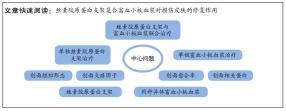
目的:观察丝素胶原蛋白支架复合富血小板血浆在皮肤创面愈合中的作用。
方法:分别制备丝素胶原蛋白支架、SD大鼠富血小板血浆。取8周龄SD大鼠48只,每只背部制作2个直径2 cm的全层皮肤缺损创面,分4组处理:空白组缺损处注射生理盐水,单纯支架组缺损处植入丝素胶原蛋白复合支架,富血小板血浆组创缘注射同种异体富血小板血浆,联合组缺损处植入丝素胶原蛋白复合支架+创缘注射同种异体富血小板血浆,每组12只。造模后检测创面愈合率、创面炎症因子水平、创面组织学观察及相关蛋白表达。

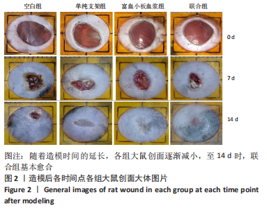
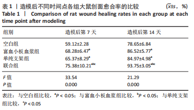
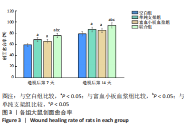
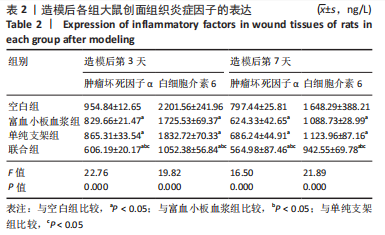
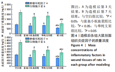
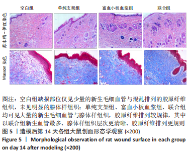
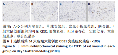
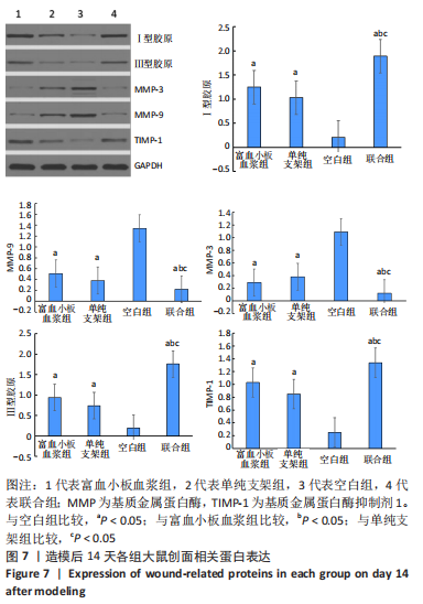
结果与结论:①联合组造模后第7,14天的创面愈合率大于空白组、单纯支架组、富血小板血浆组(P < 0.05)。②联合组造模后第7,14天的肿瘤坏死因子α、白细胞介素6水平均低于空白组、单纯支架组、富血小板血浆组(P < 0.05)。③造模后第14天的苏木精-伊红及Masson染色显示,空白组缺损处仅见少量的新生毛细血管与混乱排列的胶原纤维组织;其他3组可见大量的新生毛细血管与腺体样组织,胶原纤维排列较规律,其中以联合组新生血管最多、腺体样组织层次更清晰、胶原纤维排列更规则。免疫组化染色显示,空白组、单纯支架组、富血小板血浆组CD31+细胞密度少于联合组(P < 0.05)。④Western blot检测显示,相较于空白组、单纯支架组、富血小板血浆组,联合组创面Ⅰ型胶原、Ⅲ型胶原、基质金属蛋白酶抑制剂1的蛋白表达升高(P < 0.05),基质金属蛋白酶3、基质金属蛋白酶9蛋白表达降低(P < 0.05)。⑤结果表明,丝素胶原蛋白支架复合富血小板血浆可通过抑制炎症反应、增加微血管密度、调节细胞外基质的代谢平衡来促进皮肤创面愈合。

https://orcid.org/0000-0002-2921-6046(刘继超)

中国组织工程研究杂志出版内容重点:生物材料;骨生物材料口腔生物材料纳米材料缓释材料材料相容性组织工程


关键词: 丝素胶原蛋白复合支架, 富血小板血浆, 全层皮肤缺损, 皮肤损伤, 创面, 创面愈合

Abstract: BACKGROUND: Tissue engineering scaffolds constructed by collagen and silk fibroin are widely used in tissue engineering fields such as skin, nerves, blood vessels, bone, and cartilage. Platelet-rich plasma is a platelet concentrate obtained by centrifuging blood twice. It contains a variety of growth factors required for tissue repair and can promote tissue regeneration and wound healing. 
OBJECTIVE: To observe the effect of silk fibroin collagen scaffold combined with platelet-rich plasma on skin wound healing. 
METHODS: Silk fibroin collagen scaffolds and SD rat platelet-rich plasma were prepared separately. Totally 48 8-week-old SD rats were selected, and 2 full-thickness skin defect wounds with a diameter of 2 cm were made on the back of each rat. They were randomly divided into four groups (n=12). In the blank group, normal saline was injected into the defect site. Silk fibroin-collagen composite scaffolds were implanted in the defects of the simple scaffold group. The platelet-rich plasma group was injected with allogeneic platelet-rich plasma at the wound margin. In the combined group, silk fibroin-collagen composite scaffolds were implanted in the defect site and allogeneic platelet-rich plasma was injected into the wound margin. Wound healing rate, wound inflammatory factor level, wound histological morphology and related protein expression were detected after model establishment.
RESULTS AND CONCLUSION: (1) The wound healing rate of the combined group on days 7 and 14 after modeling was higher than that of the blank group, the simple scaffold group, and the platelet-rich plasma group (P < 0.05). (2) The levels of tumor necrosis factor α and interleukin-6 in the combined group on days 7 and 14 days after modeling were lower than those in the blank group, the simple scaffold group, and the platelet-rich plasma group (P < 0.05). (3) On day 14 after modeling, hematoxylin-eosin staining and Masson staining showed that only a few new capillaries and disordered collagen fiber tissue were seen at the defect site of the blank group. A large number of new capillaries and glandular tissue were observed in the other three groups. Collagen fibers were arranged more regularly, among which the combined group had the most new blood vessels, the glandular tissue layers were clearer, and the collagen fibers were arranged more regularly. Immunohistochemical staining displayed that the density of CD31+ cells in blank group, simple scaffold group and platelet-rich plasma group was lower than that in combined group (P < 0.05). (4) Western blot assay results showed that, compared with the blank group, the simple scaffold group, and the platelet-rich plasma group, the protein expression levels of type I collagen, type III collagen, and matrix metalloproteinase inhibitor 1 in the wound were increased in the combined group (P < 0.05), and the protein expression levels of matrix metalloproteinase 3 and matrix metalloproteinase 9 decreased (P < 0.05). (5) It is concluded that fibroin collagen scaffold combined with platelet-rich plasma can promote wound healing by inhibiting inflammatory response, increasing microvascular density and regulating the metabolic balance of extracellular matrix.

Key words: silk fibroin collagen composite scaffold, platelet-rich plasma, full-thickness skin defect, skin injury, wound surface, wound healing

中图分类号: