中国组织工程研究 ›› 2022, Vol. 26 ›› Issue (25): 3986-3992.doi: 10.12307/2022.403

• 干细胞移植 stem cell transplantation • 上一篇    下一篇

早孕蜕膜间充质干细胞联合丹参酮ⅡA治疗大鼠宫腔粘连

徐炜均1,胡向丹2,余冬青2   

  1. 1广州中医药大学第二临床医学院,广东省广州市   510000;2广东省中医院妇科,广东省广州市   510000
  • 收稿日期:2021-06-09 接受日期:2021-08-09 出版日期:2022-09-08 发布日期:2022-01-25
  • 通讯作者: 胡向丹,博士,主任医师,广东省中医院妇科,广东省广州市 510000
  • 作者简介:徐炜均,男,1995年生,广东省河源市人,汉族,2021年广州中医药大学毕业,硕士,主要从事中西医治疗宫腔粘连的相关研究。
  • 基金资助:
    广东省科技厅援疆项目(2017E020247008),项目负责人:胡向丹

First-trimester human decidual mesenchymal stem cells combined with tanshinone IIA in the treatment of intrauterine adhesions in rats

Xu Weijun1, Hu Xiangdan2, Yu Dongqing2   

  1. 1Second Clinical Medical College of Guangzhou University of Traditional Chinese Medicine, Guangzhou 510000, Guangdong Province, China; 2Department of Gynecology, Guangdong Province Hospital of Traditional Chinese Medicine, Guangzhou 510000, Guangdong Province, China
  • Received:2021-06-09 Accepted:2021-08-09 Online:2022-09-08 Published:2022-01-25
  • Contact: Hu Xiangdan, MD, Chief physician, Department of Gynecology, Guangdong Province Hospital of Traditional Chinese Medicine, Guangzhou 510000, Guangdong Province, China
  • About author:Xu Weijun, Master, Second Clinical Medical College of Guangzhou University of Traditional Chinese Medicine, Guangzhou 510000, Guangdong Province, China
  • Supported by:
    Aid Xinjiang Project of Guangdong Provincial Department of Science and Technology, No. 2017E020247008 (to HXD)

摘要:

文题释义:
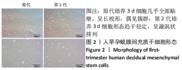
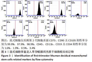
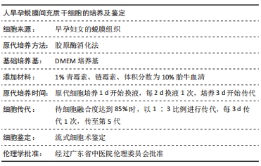
人早孕蜕膜间充质干细胞:来源于正常的子宫蜕膜组织,在胎盘胎膜发育过程中可分泌多种蛋白,并随着孕周增大与绒毛膜细胞共同发育为胎盘,具有强大的增殖能力。该研究采用胶原酶消化法分离出人早孕蜕膜间充质干细胞,流式细胞仪检测可表达间充质干细胞表面标志物CD90、CD73和CD105,不表达造血干细胞表面标志物CD34 和CD45。
丹参酮ⅡA:丹参酮是从中药丹参(唇形科植物丹参Salvia miltiorrhiza Bunge根)中提取的具有抑菌作用的脂溶性菲醌化合物,从中分得丹参酮Ⅰ、丹参酮ⅡA、丹参酮ⅡB、隐丹参酮、异隐丹参酮等10余个丹参酮单体。丹参酮Ⅱ是丹参中脂溶性成分的代表,集中分布在丹参根的皮部,常作为肿瘤、心血管疾病、炎症、动脉粥样硬化、纤维化等疾病的治疗用药。

背景:人早孕蜕膜间充质干细胞及丹参酮ⅡA具有抗粘连作用,均对宫腔粘连有一定疗效,但其具体机制待进一步研究。
目的:探索人早孕蜕膜间充质干细胞联合丹参酮ⅡA治疗宫腔粘连的效果及机制。
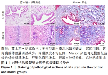
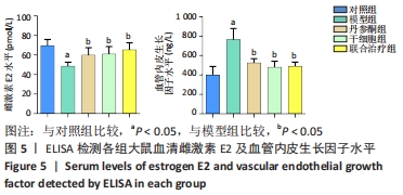
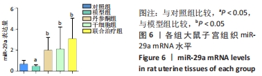
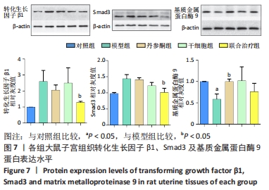
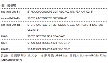
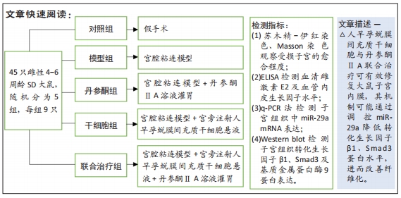
方法:采用胶原酶Ⅰ消化法分离培养人早孕蜕膜间充质干细胞,选取45只雌性4-6周龄SD大鼠,随机分为5组,每组9只,分别为对照组、模型组、丹参酮组、干细胞组及联合治疗组。对照组进行假手术;模型组进行机械刮宫造模后不干预;干细胞组则在造模后7 d经宫旁注射0.2 mL人早孕蜕膜间充质干细胞悬液(1×10⁹ L-1),每隔3 d注射1次,共3次;丹参酮组在造模后7 d灌胃11 mg/(kg·d)丹参酮ⅡA溶液,持续灌胃1周;联合治疗组则为上述两种治疗方式的联合运用。对照组及模型组在术后5,10,15 d取材,各治疗组则在最后一次治疗后5,10,15 d后取材,苏木精-伊红染色、Masson染色观察受损子宫的愈合程度;ELISA检测血清雌激素E2及血管内皮生长因子水平;q-PCR法检测子宫组织中miR-29a mRNA表达;Western blot检测子宫组织转化生长因子β1、Smad3及基质金属蛋白酶9蛋白表达。
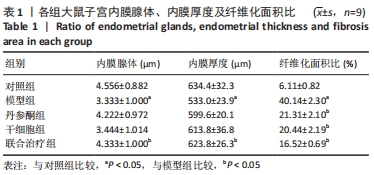
结果与结论:①与对照组比较,模型组大鼠子宫内膜纤维化面积比显著增加(P < 0.05),而内膜厚度及腺体数量显著下降(P < 0.05);与模型组比较,联合治疗组大鼠子宫内膜纤维化面积比显著下降(P < 0.05),而内膜厚度及腺体数量显著增加(P < 0.05),干细胞组及丹参酮组大鼠子宫内膜纤维化面积比也显著下降(P < 0.05);②与模型组比较,丹参酮组、干细胞组及联合治疗组血清雌激素E2水平显著升高(P < 0.05),血管内皮生长因子水平显著下降(P < 0.05),miR-29a mRNA表达显著升高(P < 0.05),联合治疗组转化生长因子β1、Smad3蛋白表达明显下降(P < 0.05),丹参酮组基质金属蛋白酶9蛋白表达明显上升(P < 0.05);③结果表明,人早孕蜕膜间充质干细胞与丹参酮ⅡA联合治疗可有效修复大鼠子宫内膜,其机制可能通过调控miR-29a降低转化生长因子β1、Smad3蛋白水平,进而改善纤维化。
缩略语:人早孕蜕膜间充质干细胞:human decidual mesenchymal stem cells,hDMSCs

https://orcid.org/0000-0001-8956-6622 (徐炜均)

中国组织工程研究杂志出版内容重点:干细胞;骨髓干细胞;造血干细胞;脂肪干细胞;肿瘤干细胞;胚胎干细胞;脐带脐血干细胞;干细胞诱导;干细胞分化;组织工程

关键词: 宫腔粘连, 早孕蜕膜干细胞, 丹参酮ⅡA, miR-29a, 转化生长因子β1, Smad3

Abstract: BACKGROUND: First-trimester human decidual mesenchymal stem cells and tanshinone IIA have anti-adhesion effect, and both have a certain effect on intrauterine adhesion, but the specific mechanism needs to be further studied.  
OBJECTIVE: To explore the efficacy and mechanism of first-trimester human decidual mesenchymal stem cells combined with tanshinone IIA in the treatment of intrauterine adhesions.
METHODS:  First-trimester human decidual mesenchymal stem cells were isolated and cultured by collagenase I digestion. A total of 45 female SD rats aged 4-6 weeks were randomly divided into 5 groups with 9 rats in each group: control group, model group, tanshinone group, stem cell group and combined treatment group. Sham operation was performed in the control group, and no intervention was performed in the model group after modeling. In the stem cell group, 0.2 mL human decidual mesenchymal stem cell suspension (1×10⁹ L-1) was injected parauterine 7 days after modeling, once every other 3 days, for a total of 3 times. In the tanshinone group, tanshinone IIA solution at 11 mg/(kg·d) was given via intragastulation for 1 week. The combined treatment group received the combination of the above two treatment methods. In the control group and model group, samples were obtained at 5, 10, and 15 days after operation and sham operation. In each treatment group, samples were taken at 5, 10, and 15 days after the last treatment. Hematoxylin-eosin staining and Masson staining were performed to observe the healing degree of the damaged uterus. Serum vascular endothelial growth factor and E2 levels were detected by ELISA. q-PCR was used to detect the expression of miR-29a mRNA in uterine tissue. The protein expression levels of transforming growth factor β1, Smad3 and matrix metalloproteinase 9 were detected by western blot assay.  
RESULTS AND CONCLUSION: (1) Compared with the control group, the endometrial fibrosis area ratio in the model group increased significantly (P < 0.05), while the endometrial thickness and the number of glands decreased significantly (P < 0.05). Compared with the model group, the degree of endometrial fibrosis in the combined treatment group was significantly lower (P < 0.05) and the thickness of the intima and the number of glands increased significantly (P < 0.05). The endometrial fibrosis area ratio of rats in the stem cell group and the tanshinone group also decreased significantly (P < 0.05). (2) Compared with the model group, the serum estrogen E2 level of the tanshinone, stem cell and the combined treatment groups was significantly increased (P < 0.05); vascular endothelial growth factor level was significantly decreased (P < 0.05); the expression of miR-29a mRNA was significantly increased (P < 0.05); the expression of transforming growth factor β1 and Smad3 protein in the combined treatment group was significantly decreased (P < 0.05), and the expression of matrix metalloproteinase 9 protein in the tanshinone group was significantly increased (P < 0.05). (3) Results suggest that the combined treatment of first-trimester human decidual mesenchymal stem cells and tanshinone IIA can effectively repair the endometrium in rats, and the mechanism may be through the regulation of miR-29a to reduce the levels of transforming growth factor β1 and Smad3 protein, and then improve the fibrosis.

Key words: intrauterine adhesion, first-trimester human decidual mesenchymal stem cells, tanshinone IIA, miR-29a, transforming growth factor-β1, Smad3

中图分类号: