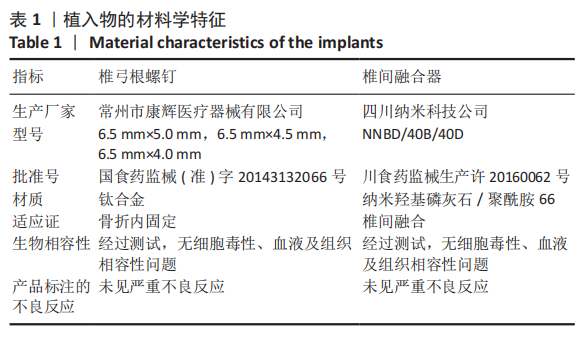
中国组织工程研究 ›› 2021, Vol. 25 ›› Issue (36): 5821-5826.doi: 10.12307/2021.349

• 脊柱植入物 spinal implant • 上一篇    下一篇

椎间自体骨植骨面积比与椎间融合率的关系

唐  强,钟德君,王  清,廖晔晖,唐  超,马  飞   

  1. 西南医科大学附属医院骨科,四川省泸州市   646000
  • 收稿日期:2021-01-11 修回日期:2021-01-12 接受日期:2021-02-27 出版日期:2021-12-28 发布日期:2021-09-17
  • 通讯作者: 钟德君,博士,主任医师,西南医科大学附属医院骨科,四川省泸州市 646000
  • 作者简介:唐强,男,1987年生,四川省泸州市人,2017年西南医科大学毕业,硕士,医师,主要从事脊柱外科方面的研究。
  • 基金资助:
    西南医科大学基金项目(2020ZRQNB041),项目负责人:唐强

Relationship between area ratio of interbody autograft and intervertebral fusion rate

Tang Qiang, Zhong Dejun, Wang Qing, Liao Yehui, Tang Chao, Ma Fei   

  1. Department of Orthopedics, Affiliated Hospital of Southwest Medical University, Luzhou 646000, Sichuan Province, China
  • Received:2021-01-11 Revised:2021-01-12 Accepted:2021-02-27 Online:2021-12-28 Published:2021-09-17
  • Contact: Zhong Dejun, MD, Chief physician, Department of Orthopedics, Affiliated Hospital of Southwest Medical University, Luzhou 646000, Sichuan Province, China
  • About author:Tang Qiang, Master, Physician, Department of Orthopedics, Affiliated Hospital of Southwest Medical University, Luzhou 646000, Sichuan Province, China
  • Supported by:
    a grant from southwest medical university, No. 2020ZRQNB041 (to TQ)

摘要:


文题释义:

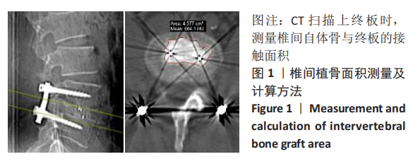
椎间自体骨植骨面积比:融合节段椎间自体骨植骨与上、下终板接触的横截面积与之相应接触终板面积的比值×100%,取其均值。

背景:目前相关报道认为椎间植骨体积是影响椎间融合率的因素之一,但相同植骨体积因个体差异导致椎间植骨与终板的接触面积不同,椎间植骨与终板的接触面积对椎间融合率的影响鲜有分析研究。
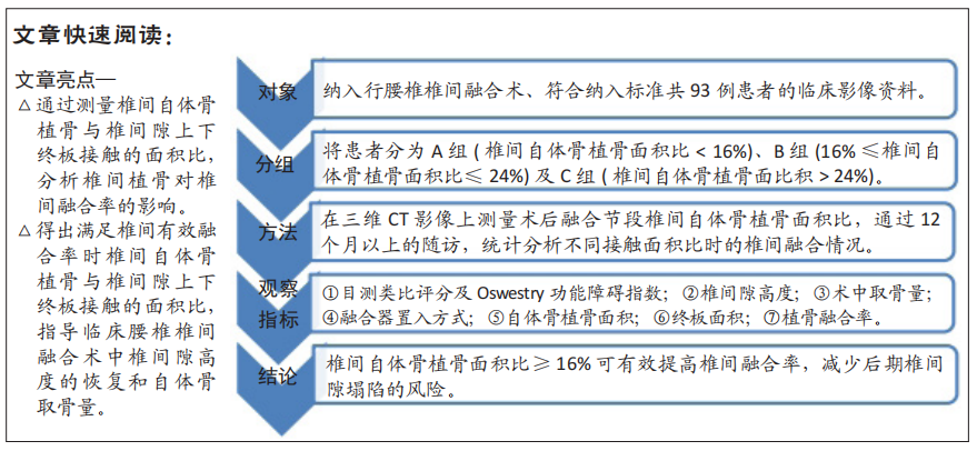
目的:探讨行腰椎后路椎间融合时自体骨植骨面积比对椎间融合率的影响。
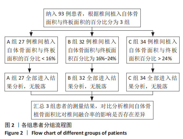
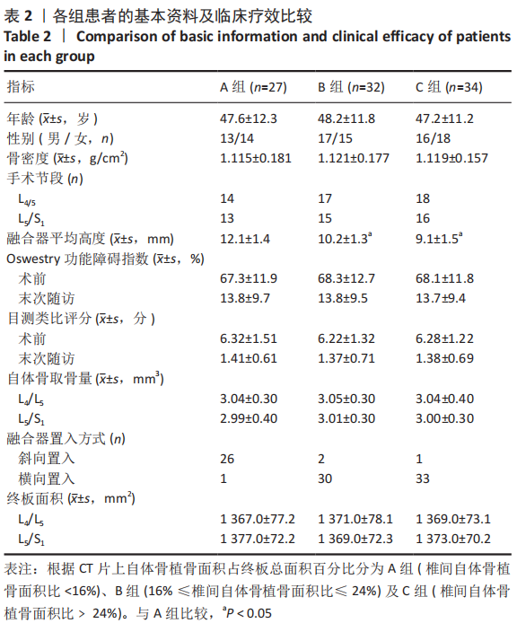
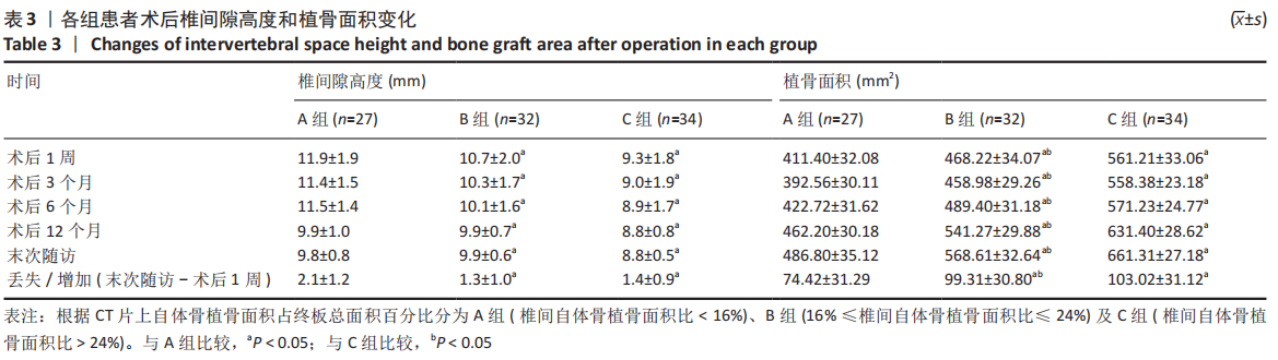
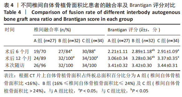
方法:回顾性分析2017年1月至2018年12月在西南医科大学附属医院脊柱外科行腰椎后路椎体间融合手术且符合纳入标准共93例患者的临床资料,记录患者术前椎间隙高度、上下终板面积、Oswestry功能障碍指数及目测类比评分、术中使用Cage大小及自体骨取骨量,术后测量自体骨植骨面积(椎间与上下终板接触的自体骨面积),取其均值;根据术后1周测量的自体骨植骨面积占终板总面积百分比分为A组(椎间自体骨植骨面积比< 16%)、B组(16%≤椎间自体骨植骨面积比≤24%)及C组(椎间自体骨植骨面积比> 24%)。术后6,12 个月及末次随访行X射线片及CT检查,观察3组患者术中融合器置入方向,记录随访时椎间隙高度、椎间自体骨植骨面积比、椎间融合Brantigan评分、Oswestry功能障碍指数及目测类比评分。测量由2名脊柱外科医师分别完成,间隔1周重复测量,检验观察者间及观察者内信度。比较分析椎间自体骨植骨面积对椎间融合率的影响。

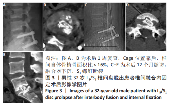
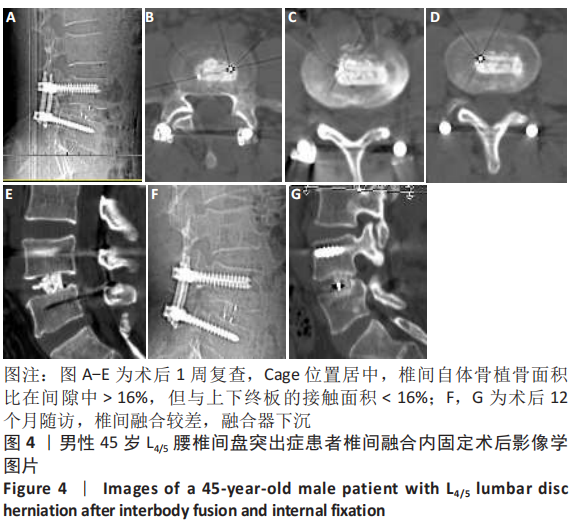
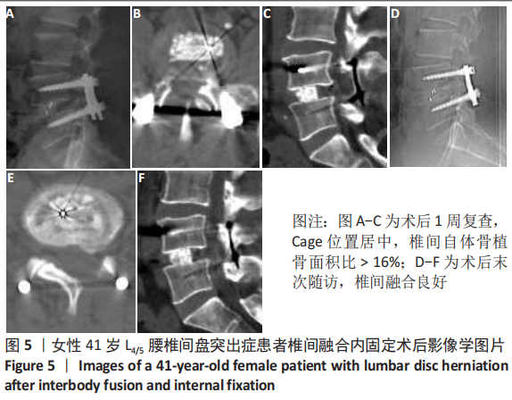
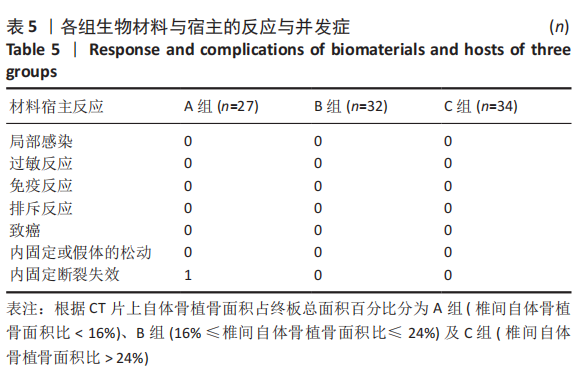
结果与结论:①椎间植骨面积测量结果观察者间及观察者内信度分别为0.924和0.913;②所有患者均获得12个月以上随访,3组患者年龄、性别及手术节段差异均无显著性意义(P > 0.05),术前椎间隙高度、Oswestry功能障碍指数及目测类比评分组间比较差异无显著性意义(P > 0.05);③A组患者27例,其中融合器斜向置入26例,横向置入1例;B组患者32例,其中融合器斜向置入2例,横向置入30例;C组患者34例,斜向置入1例,横向置入33例;④3组患者术中自体骨取骨量比较差异无显著性意义(P > 0.05);术后1周椎间隙平均高度A组大于B、C组(P=0.022,P=0.000),自体骨植骨面积A组小于B、C组(P=0.000,P=0.000);⑤术后6个月及12个月,椎间融合率A组(70.4%,88.9%)低于B组(84.4%,100%)和C组(88.2%,100%),Brantigan评分A组低于B组(P=0.027,P=0.020)和C组(P=0.018,P=0.001);⑥末次随访3组患者Oswestry功能障碍指数及目测类比评分比较差异均无显著性意义(P > 0.314),椎间隙高度丢失A组大于B、C组(P=0.007,P=0.013),椎间植骨融合成骨增加面积A组低于B、C组(P=0.003,P=0.000),3组融合率及Brantigan评分比较差异无显著性意义(P=0.902,P=0.712);⑦提示椎间自体骨植骨面积比≥16%可有效提高椎间融合率,减少后期椎间隙塌陷的风险。

https://orcid.org/0000-0002-3776-9695 (唐强) 

中国组织工程研究杂志出版内容重点:人工关节;骨植入物;脊柱;骨折;内固定;数字化骨科;组织工程

关键词: 腰椎, 后路椎体间融合, 自体骨, 植骨面积, 融合率, 单侧椎板间开窗

Abstract: BACKGROUND: At present, it is considered that the volume of intervertebral bone graft is one of the factors affecting the intervertebral fusion rate, but the same volume of intervertebral bone graft leads to different release area of intervertebral bone graft and endplate due to individual differences, and the impact of release area of intervertebral bone graft and endplate on the intervertebral fusion rate is rarely analyzed.  
OBJECTIVE: To investigate the effect of area ratio of interbody autograft on intervertebral fusion rate during posterior lumbar interbody fusion.
METHODS:  Clinical data of 93 patients who underwent posterior lumbar interbody fusion in Department of Spinal Surgery, Affiliated Hospital of Southwest Medical University from January 2017 to December 2018 were retrospectively analyzed. The preoperative intervertebral space height, upper and lower endplate area, Oswestry disability index and visual analogue scale score, Cage size and the amount of autologous bone, and area of autologous bone graft (the area of autologous bone contacting the upper and lower endplates) were recorded, and the average value was calculated. According to the percentage of autogenous bone graft area in the total endplate area measured one week after operation, they were divided into group A (area ratio of interbody autograft < 16%), group B (16% ≤ area ratio of interbody autograft ≤ 24%) and group C (area ratio of interbody autograft > 24%). X-ray and CT examinations were performed at 6 and 12 months and the last follow-up. The direction of cage placement was observed in the three groups. The height of intervertebral space, the area ratio of intervertebral autograft bone graft, brantigan score, Oswestry disability index and visual analogue scale score were recorded at the follow-up. The measurement was performed by two spine surgeons, and repeated at an interval of one week to test inter observer and intra observer reliability. The effect of the area of autologous bone graft on the intervertebral fusion rate was compared and analyzed.  
RESULTS AND CONCLUSION: (1) The inter observer and intra observer reliabilities were 0.924 and 0.913, respectively. (2) All patients were followed up for more than 12 months. There were no significant differences in age, gender and surgical segment among the three groups (P > 0.05). There were no significant differences in preoperative intervertebral height, Oswestry disability index and visual analogue scale score among the three groups (P > 0.05). (3) There were 27 cases in group A, including 26 cases of oblique placement and 1 case of transverse placement, 32 cases in group B, including 2 cases of oblique placement and 30 cases of transverse placement, 34 cases in group C, 1 case of oblique placement and 33 cases of transverse placement. (4) There was no significant difference in the amount of intraoperative autologous bone extraction among the three groups (P > 0.05), and the average height of intervertebral space in group A was higher than that in group B and group C one week after operation (P=0.022, P=0.00). The area of autogenous bone graft in group A was smaller than that in group B and group C (P=0.000, P=0.000). (5) At 6 and 12 months after operation, the fusion rate in group A (70.4%, 88.9%) was lower than that in group B (84.4%, 100%) and group C (88.2%, 100%), and the Brantigan score in group A was lower than that in group B (P=0.027, P=0.020) and group C (P=0.018, P=0.001). (6) At the last follow-up, there was no significant difference in Oswestry disability index and visual analogue scale score among the three groups (P > 0.314). The height loss of intervertebral space in group A was greater than that in group B and group C (P=0.007, P=0.013). The increased area of intervertebral fusion in group A was lower than that in group B and group C (P=0.003, P=0.000). There was no significant difference in fusion rate and Brantigan score among groups A, B and C (P=0.902, P=0.712). (7) It is suggested that the percentage of an area ratio of interbody autograft in the total endplate area greater than or equal to 16% can effectively improve the interbody fusion rate and reduce the risk of late intervertebral space collapse.

Key words: lumbar spine, posterior interbody fusion, autogenous bone, bone graft area, fusion rate, unilateral interlaminar fenestration

中图分类号: