中国组织工程研究 ›› 2023, Vol. 27 ›› Issue (10): 1507-1513.doi: 10.12307/2023.369

• 骨髓干细胞 bone marrow stem cells • 上一篇    下一篇

解整合素金属蛋白酶10与激素性股骨头坏死患者骨髓间充质干细胞的增殖及成骨分化

樊  俊1,吴陈欢2,程中华2   

  1. 1长江大学,湖北省荆州市 434023;2长江大学附属黄冈市中心医院骨科II,湖北省黄冈市 438000
  • 收稿日期:2022-04-13 接受日期:2022-06-27 出版日期:2023-04-08 发布日期:2022-09-06
  • 通讯作者: 程中华,硕士,主任医师,长江大学附属黄冈市中心医院骨科II,湖北省黄冈市 438000
  • 作者简介:樊俊,男,1991年生,湖北省黄冈市人,汉族,长江大学在读硕士,主要从事骨与关节损伤方向的研究。
  • 基金资助:
    湖北省自然科学基金(s2015-01-01330084),项目负责人:程中华

Effect of a disintegrin and metalloproteases 10 on proliferation and osteogenic differentiation of bone marrow mesenchymal stem cells induced by steroid-induced osteonecrosis of femoral head

Fan Jun1, Wu Chenhuan2, Cheng Zhonghua2   

  1. 1Yangtze University, Jingzhou 434023, Hubei Province, China; 2Department of Orthopedics II, Huanggang Central Hospital Affiliated to Yangtze University, Huanggang 438000, Hubei Province, China
  • Received:2022-04-13 Accepted:2022-06-27 Online:2023-04-08 Published:2022-09-06
  • Contact: Cheng Zhonghua, Master, Chief physician, Department of Orthopedics II, Huanggang Central Hospital Affiliated to Yangtze University, Huanggang 438000, Hubei Province, China
  • About author:Fan Jun, Master candidate, Yangtze University, Jingzhou 434023, Hubei Province, China
  • Supported by:
    Natural Science Foundation of Hubei Province, No. s2015-01-01330084 (to CZH)

摘要:

文题释义:
解整合素金属蛋白酶10:由金属蛋白酶、富含半胱氨酸、分解素和表皮生长因子样结构域组成的模块蛋白,可参与多种生物学功能,包括炎症、凋亡、细胞黏附、细胞代谢、肿瘤转移和自身免疫等。
激素性股骨头坏死:长时间或大量使用激素可导致局部骨内压力增加、血液循环紊乱,从而引起骨细胞进一步缺血、坏死和骨小梁断裂,最终导致股骨头塌陷的一类骨骼疾病。

背景:众多研究发现,激素性股骨头坏死的病理进展与骨髓间充质干细胞增殖及成骨分化异常有关,其中解整合素金属蛋白酶10(A disintegrin and metalloproteases 10,ADAM10)高度参与此过程,但具体作用机制尚不清楚。
目的:探讨ADAM10对激素性股骨头坏死患者来源骨髓间充质干细胞增殖及成骨分化的影响及其可能机制。
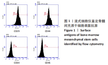
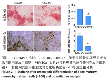
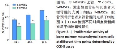
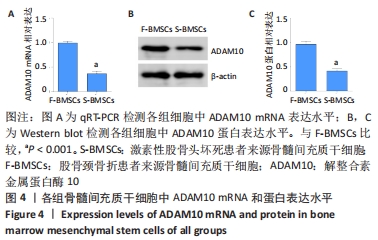
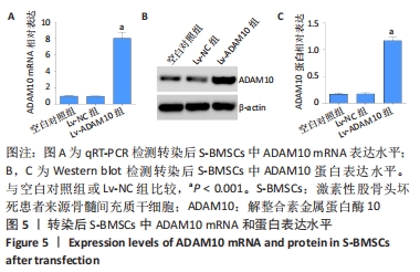
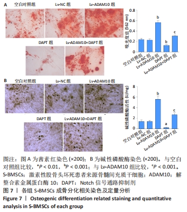
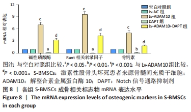
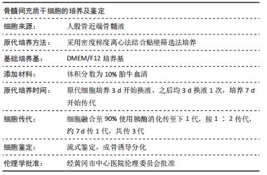
方法:选择23例激素性股骨头坏死患者和17例股骨颈骨折患者,于术中无菌条件下取股骨近端骨髓,采用密度梯度离心法分离骨髓间充质干细胞,分别得到激素性股骨头坏死患者来源骨髓间充质干细胞(S-BMSCs)和股骨颈骨折患者来源骨髓间充质干细胞(F-BMSCs),体外培养至第3代,采用CCK-8检测两组细胞增殖活性;茜素红染色和碱性磷酸酶染色观察两组细胞成骨分化能力;qRT-PCR和Western blot检测两组细胞中ADAM10 mRNA和蛋白表达水平。采用ADAM10过表达载体慢病毒感染S-BMSCs,再联合Notch信号通路抑制剂DAPT进行处理,CCK-8检测细胞增殖活性;茜素红染色和碱性磷酸酶染色观察细胞成骨分化能力;qRT-PCR检测细胞中成骨标志物碱性磷酸酶、Runt相关转录因子2、骨钙素mRNA表达水平;Western blot检测细胞中Notch信号通路相关蛋白Notch1、NICD、Hes1表达水平。
结果与结论:①S-BMSCs的增殖活性、成骨分化能力及ADAM10表达水平均显著低于F-BMSCs(均P < 0.01);②ADAM10过表达后S-BMSCs增殖活性、成骨分化能力及碱性磷酸酶、Runt相关转录因子2、骨钙素mRNA表达水平和Notch1、NICD、Hes1蛋白表达水平均显著升高(P < 0.05);然而,DAPT联合干预可显著抑制ADAM10过表达对S-BMSCs增殖活性和成骨分化能力的促进作用,并抑制Notch信号通路的活化;③结果表明,ADAM10在S-BMSCs中低表达,ADAM10过表达可增强S-BMSCs的增殖活性及成骨分化能力,其作用机制可能与激活Notch信号通路有关。

https://orcid.org/0000-0001-5382-7747 (樊俊) 

中国组织工程研究杂志出版内容重点:干细胞;骨髓干细胞;造血干细胞;脂肪干细胞;肿瘤干细胞;胚胎干细胞;脐带脐血干细胞;干细胞诱导;干细胞分化;组织工程

关键词: 激素性股骨头坏死, 骨髓间充质干细胞, 解整合素金属蛋白酶10, Notch信号通路, 成骨分化

Abstract: BACKGROUND: Many studies have found that the pathological progression of steroid-induced osteonecrosis of the femoral head is related to abnormal proliferation and osteogenic differentiation of bone marrow mesenchymal stem cells, in which a disintegrin and metalloproteases 10 (ADAM10) is highly involved, but the specific mechanism remains unclear.  
OBJECTIVE: To investigate the effects of ADAM10 on proliferation and osteogenic differentiation of bone marrow mesenchymal stem cells derived from steroid-induced osteonecrosis of the femoral head patients and its possible mechanism.
METHODS:  Bone marrow was harvested from proximal femur in 23 patients with steroid-induced osteonecrosis of the femoral head and 17 patients with femoral neck fracture under sterile conditions. Bone marrow mesenchymal stem cells from steroid-induced osteonecrosis of the femoral head (S-BMSCs) and femoral neck fracture (F-BMSCs) were isolated by density gradient centrifugation, and cultured to the third generation in vitro. The proliferation activity of cells in the two groups was detected by CCK-8 assay. Alizarin red staining and alkaline phosphatase staining were used to observe the osteogenic differentiation ability of cells in the two groups. qRT-PCR and western blot assay were used to detect the levels of ADAM10 mRNA and protein in cells of the two groups. S-BMSCs were infected with ADAM10 overexpression lentivirus vector, and then treated with Notch signaling pathway inhibitor DAPT. The proliferation activity of these cells was detected by CCK-8 assay. Alizarin red staining and alkaline phosphatase staining were used to observe the osteogenic differentiation of cells. qRT-PCR was used to detect the expression levels of osteogenic markers alkaline phosphatase, Runt-related transcription factor 2, and osteocalcin mRNA. The expression levels of Notch signaling pathway related proteins Notch1, NICD, and Hes1 were detected by western blot assay.  
RESULTS AND CONCLUSION: (1) The proliferation activity, osteogenic differentiation ability and the expression level of ADAM10 in S-BMSCs were significantly lower than those in F-BMSCs (P < 0.01). (2) After ADAM10 overexpression, the proliferation activity, osteogenic differentiation ability, the expression levels of alkaline phosphatase, Runt-related transcription factor 2, and osteocalcin mRNA and Notch1, NICD and Hes1 protein of S-BMSCs were significantly increased (P < 0.05). However, combined intervention with DAPT significantly inhibited the promoting effects of ADAM10 overexpression on proliferation activity and osteogenic differentiation ability of S-BMSCs, and inhibited the activation of Notch signaling pathway. (3) The results show that ADAM10 can be underexpressed in S-BMSCs, and its overexpression can enhance the proliferation and osteogenic differentiation ability of S-BMSCs, and its mechanism may be related to the activation of Notch signaling pathway.

Key words: steroid-induced osteonecrosis of femoral head, bone marrow mesenchymal stem cell, a disintegrin and metalloproteases 10, Notch signaling pathway, osteogenic differentiation

中图分类号: