[1] ZHANG X, ZHANG C, LIN Y, et al. Nanocomposite Membranes Enhance Bone Regeneration Through Restoring Physiological Electric Microenvironment. ACS Nano. 2016;10(8):7279-7286.
[2] LEPPIK L, OLIVEIRA KMC, BHAVSAR MB, et al. Electrical stimulation in bone tissue engineering treatment. Eur J Trauma Emerg Surg. 2020;46(2):231-244.
[3] 朱子馨,周俊余,苟雪.可拉伸的钛酸钡压电膜上细胞黏附行为的研究[J].医用生物力学,2021,36(S1):164.
[4] 熊莹,许燕,周建平,等.组织工程研究中的电活性生物材料[J].中国组织工程研究,2019,23(34):5523-5530.
[5] ZHENG T, HUANG Y, ZHANG X, et al. Mimicking the electrophysiological microenvironment of bone tissue using electroactive materials to promote its regeneration. J Mater Chem B. 2020;8(45):10221-10256.
[6] KHARE D, BASU B, DUBEY AK. Electrical stimuLation and piezoelectric biomaterials for bone tissue engineering applications. Biomaterials. 2020;258:120280.
[7] TANDON B, BLAKER JJ, CARTMELL SH. Piezoelectric materials as stimulatory biomedical materials and scaffolds for bone repair. Acta Biomater. 2018;73:1-20.
[8] KAO FC, CHIU PY, TSAI TT, et al. The application of nanogenerators and piezoelectricity in osteogenesis. Sci Technol Adv Mater. 2019;20(1):1103-1117.
[9] TANG Y, WU C, WU Z, et al. Fabrication and in vitro biological properties of piezoelectric bioceramics for bone regeneration. Sci Rep. 2017;7:43360.
[10] 胡龙.HA/BaTiO_3压电仿生骨植入材料的制备及性能研究[D].西安:西安理工大学,2014.
[11] LIU W, LI X, JIAO Y, et al. Biological Effects of a Three-Dimensionally Printed Ti6Al4V Scaffold Coated with Piezoelectric BaTiO3 Nanoparticles on Bone Formation. ACS Appl Mater Interfaces. 2020;12(46):51885-51903.
[12] EHTERAMI A, KAZEMI M, NAZARI B, et al. Fabrication and characterization of highly porous barium titanate based scaffold coated by Gel/HA nanocomposite with high piezoelectric coefficient for bone tissue engineering applications. J Mech Behav Biomed Mater. 2018;79:195-202.
[13] LI Y, LIAO C, TJONG SC. Electrospun Polyvinylidene Fluoride-Based Fibrous Scaffolds with Piezoelectric Characteristics for Bone and Neural Tissue Engineering. Nanomaterials (Basel). 2019;9(7):952.
[14] AMARO L, CORREIA DM, MARQUES-ALMEIDA T, et al. Tailored Biodegradable and Electroactive Poly(Hydroxybutyrate-Co-Hydroxyvalerate) Based Morphologies for Tissue Engineering Applications. Int J Mol Sci. 2018;19(8):2149.
[15] AMARO L, CORREIA DM, MARTINS PM, et al. Morphology Dependence Degradation of Electro- and Magnetoactive Poly(3-hydroxybutyrate-co-hydroxyvalerate) for Tissue Engineering Applications. Polymers (Basel). 2020; 12(4):953.
[16] 郭莉莉.聚L-乳酸基驻极体膜的制备及其性能研究[D].郑州:郑州大学,2013.
[17] LI Y, DAI X, BAI Y, et al. Electroactive BaTiO3 nanoparticle-functionalized fibrous scaffolds enhance osteogenic differentiation of mesenchymal stem cells. Int J Nanomedicine. 2017;12:4007-4018.
[18] 段瑞侠,陈金周,刘文涛,等.聚乳酸基压电材料的研究和应用[J/OL].材料导报:1-18[2021-11-17].http://kns.cnki.net/kcms/detail/50.1078.TB. 20211115.1801.037.html.
[19] SAEIDI B, DERAKHSHANDEH MR, CHERMAHINI MD, et al. Novel Porous Barium Titanate/Nano-Bioactive Glass Composite with High Piezoelectric Coefficient for Bone Regeneration Applications. J Mater Eng Perform. 2020;29(8):1-8.
[20] 魏子钦,夏翔,李勤,等.钛酸钡/硅酸钙复合生物活性压电陶瓷的制备及性能研究[J/OL].无机材料学报:1-8[2021-12-19].http://kns.cnki.net/kcms/detail/31.1363.TQ.20211118.1850.026.html.
[21] 朱子馨,周俊余,苟雪.可拉伸的钛酸钡压电膜上细胞黏附行为的研究[J].医用生物力学,2021,36(S1):164.
[22] 刘国峰.自供电刺激骨支架的激光增材制造及其性能研究[D].赣州:江西理工大学,2021.
[23] LIU J, GU H, LIU Q, et al. An intelligent material for tissue reconstruction: The piezoelectric property of polycaprolactone/barium titanate composites. Mater Lett. 2019;236(FEB.1):686-689.
[24] MANCUSO E, SHAH L, JINDAL S, et al. Additively manufactured BaTiO3 composite scaffolds: A novel strategy for load bearing bone tissue engineering applications. Mater Sci Eng C Mater Biol Appl. 2021;126:112192-112192.
[25] PROKHOROV E, BÁRCENAS GL, ESPAÑA SÁNCHEZ BL, et al. Chitosan-BaTiO3 nanostructured piezopolymer for tissue engineering. Colloids Surf B Biointerfaces. 2020;196:111296.
[26] BAGCHI A, MEKA SR, RAO BN, et al. Perovskite ceramic nanoparticles in polymer composites for augmenting bone tissue regeneration. Nanotechnology. 2014; 25(48):485101.
[27] 傅志昂.辐射法制备有机-无机杂化纳米粒子及其在高分子材料中的应用研究[D].北京:中国科学院大学(中国科学院上海应用物理研究所),2021.
[28] 尹兴,孙诚,付春英,等.纳米二氧化钛/聚乳酸抗菌薄膜的制备和性能[J].包装工程,2017,38(15):36-40.
[29] 任煜,李猛,尤祥银.驻极处理对聚乳酸熔喷材料性能的影响[J].纺织学报, 2015,36(9):13-17.
[30] 吴聪.钛表面生物压电涂层的构建及其骨修复促进机制研究[D].西安:西安理工大学,2021.
[31] 闫欢欢.贻贝仿生导电性苯胺齐聚物生物材料的制备及在组织工程中的应用[D].北京:中国科学技术大学,2020.
[32] 曹钰彬,刘畅,潘韦霖,等.引导骨再生屏障膜改良的研究进展[J].华西口腔医学杂志,2019(3):325-329.
[33] LI X, WEI L, LI J, et al. Multifunctional SDF-1-loaded hydroxyapatite/polylactic acid membranes promote cell recruitment, immunomodulation, angiogenesis, and osteogenesis for biomimetic bone regeneration. Appl Mater Today. 2021;22:100942.
[34] 封小霞,侯玮玮,金晓婷,等.构建聚乳酸-羟基乙酸电纺丝-壳聚糖电喷微球牙周仿生膜[J].中国组织工程研究,2020,24(4):511-516.
[35] 方炜.聚乳酸/生物玻璃引导骨再生膜制备及体外实验研究[D].广州:南方医科大学,2015.
[36] CHEN W, YU Z, PANG J, et al. Fabrication of Biocompatible Potassium Sodium Niobate Piezoelectric Ceramic as an Electroactive Implant. Materials (Basel). 2017;10(4):345.
[37] 田春燕.电活性生物骨修复复合材料(BT-HA)/PVDF的制备和性能研究[D].北京:北京化工大学,2009.
[38] 朱培君,赖春花,程鸣威,等.电活性生物膜通过调控巨噬细胞极化促进骨再生修复的体外研究[J].实用医学杂志,2021,37(10):1257-1262.
[39] 何逸恒,程鸣威,朱培君,等.电活性生物膜促进大鼠的体内成骨[J].中国组织工程研究,2022,26(28):4662-4667.
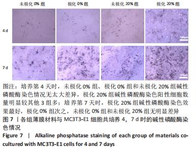
|