[1] STONE C. Cleft lip and palate: etiology, epidemiology, preventive and intervention strategies. Anat Physiol. 2013;4(3):1-5.
[2] KALASKAR R, KALASKAR A, NAQVI FS, et al. Prevalence and evaluation of environmental risk factors associated with cleft lip and palate in a central Indian population. Pediatr Dent. 2013;35(3):279-283.
[3] FAN D, WU S, LIU L, et al. Prevalence of non-syndromic orofacial clefts: based on 15, 094, 978 Chinese perinatal infants. Oncotarget. Oncotarget. 2018;9(17): 13981-13990.
[4] 孙凤霞,申铁兵.非综合征性唇腭裂流行病学的研究进展[J].口腔颌面外科杂志,2014,24(2):154-157.
[5] MOSMULLER D, MAAL TJ, PRAHL C, et al. Comparison of two- and three-dimensional assessment methods of nasolabial appearance in cleft lip and palate patients: Do the assessment methods measure the same outcome. J Craniomaxillofac Surg. 2017;45(8):1220-1226.
[6] GATTANI S, JU X, GILLGRASS T, et al. An innovative assessment of the dynamics of facial movements in surgically managed unilateral cleft lip and palate using 4D imaging. Cleft Palate Craniofac J. 2020;57(2):1-9.
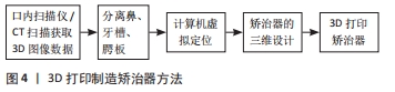
[7] 姚金凤,邓梦昭,谢添,等.口腔数字化设计在唇腭裂患者前牙美学修复中的应用研究[J].华西口腔医学杂志,2021,39(5):582-590.
[8] 李彪,姜腾飞,沈舜尧,等.3D打印个体化钛板在正颌手术中的应用及其准确性评价[J].中国口腔颌面外科杂志,2016,14(5):6.
[9] 王敏娇,蔡鸣,姜闻博,等.双侧完全性唇腭裂鼻畸形患儿唇裂术后个性化鼻模的计算机辅助设计和制作 [J].组织工程与重建外科杂志,2021,17(2):118-121.
[10] 张天嘉,王旭东.3D打印导板和个性化钛板在正颌外科合并肋骨移植术中的应用[C].第十四次中国口腔颌面外科学术会议论文汇编,2018,1(1):651-652.
[11] GIANNETTI S, BIZZOTTO N, STANCATI A, et al. Minimally invasive fixation in tibial plateau fractures using an pre-operative and intra-operative real size 3D printing. Injury. 2017;48(3):784-788.
[12] OOSTERKAMP BCM, MEER WJVD, RUTENFRANS M, et al. Reliability of linear measurements on a virtual bilateral cleft lip and palate model. Cleft Palate Craniofac J. 2006;43(5):519-523.
[13] YU Q, GONG X, WANG G M, et al. A novel technique for presurgical nasoalveolar molding using computer-aided reverse engineering and rapid prototyping. J Craniofac Surg. 2011;22(1):142-148.
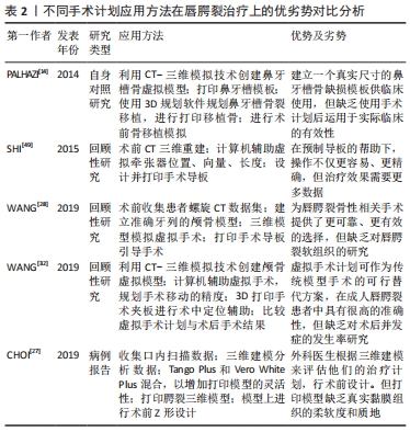
[14] PALHAZI P, NEMES B, SWENNEN G, et al. Three-dimensional simulation of the nasoalveolar cleft defect. Cleft Palate Craniofac J. 2014;51(5):593-596.
[15] ZHENG Y, LU B, ZHANG J, et al. CAD/CAM silicone simulator for teaching cheiloplasty: description of the technique. Br J Oral Maxillofac Surg. 2015;53(2): 194-196.
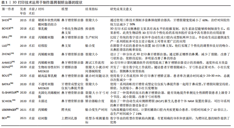
[16] SHEN C, YAO CA, MAGEE W 3rd, et al. Presurgical nasoalveolar molding for cleft lip and palate: the application of digitally designed molds. Plast Reconstr Surg. 2015;135(6):1007e-1015e.
[17] LIOUFAS PA, QUAYLE MR, LEONG JC, et al. 3D printed models of cleft palate pathology for surgical education. Plast Reconstr Surg Glob Open. 2016;4(9):e1029.
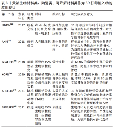
[18] HIXON KR, MELVIN AM, LIN AY, et al. Cryogel scaffolds from patient-specific 3D-printed molds for personalized tissue-engineered bone regeneration in pediatric cleft-craniofacial defects. J Biomater Appl. 2017;32(5):598-611.
[19] KASAVEN CP, MCINTYRE GT, MOSSEY PA. Accuracy of both virtual and printed 3-dimensional models for volumetric measurement of alveolar clefts before grafting with alveolar bone compared with a validated algorithm: a preliminary investigation. Br J Oral Maxillofac Surg. 2017;55(1):31-36.
[20] ALALI AB, GRIFFIN MF, CALONGE WM, et al. Evaluating the use of cleft lip and palate 3D-printed models as a teaching aid. J Surg Educ. 2018;75(1):200-208.
[21] DU F, LI B, YIN N, et al. Volumetric analysis of alveolar bone defect using three-dimensional-printed models versus computer-aided engineering. J Craniofac Surg. 2017;28(2):383-386.
[22] REIGHARD CL, GREEN K, ROONEY DM, et al. Development of a novel, low-cost, high-fidelity cleft lip repair surgical simulatorusing computer-aided design and 3-Dimensional printing. JAMA Facial Plastic Surg. 2018;12(1):77-79.
[23] NICOT R, COULY G, FERRI J, et al. Three-dimensional printed haptic model from a prenatal surface-rendered oropalatal sonographic view: a new tool in the surgical planning of cleft lip/palate. Int J Oral Maxillofac Surg. 2018;47(1):44-47.
[24] BOYER CJ, WOERNER JE, GALEA C, et al. Personalized bioactive nasal supports for postoperative cleft rhinoplasty. J Oral Maxillofac Surg. 2018;76(7):1562 e1-1562 e5.
[25] AHN G, LEE JS, YUN WS, et al. Cleft alveolus reconstruction using a three-dimensional printed bioresorbable scaffold with human bone marrow cells. J Craniofac Surg. 2018;29(7):1880-1883.
[26] LUO D, LI T, WANG H, et al. Three-dimensional printing of personalized nasal stents for patients with cleft lip. Cleft Palate Craniofac J. 2019;56(4):521-524.
[27] CHOI YS, SHIN HS. Preoperative planning and simulation in patients with cleft palate using intraoral three-dimensional scanning and printing. J Craniofac Surg. 2019;30(7):2245-2248.
[28] WANG Y, ZHANG Z, LIU Y, et al. Virtual Surgical Planning Assisted Management for Cleft-Related Maxillary Hypoplasia. J Craniofac Surg. 2019;30(6):1745-1749.
[29] ZHENG J, HE H, KUANG W, et al. Presurgical nasoalveolar molding with 3D printing for a patient with unilateral cleft lip, alveolus, and palate. Am J Orthod Dentofacial Orthop. 2019;156(3):412-419.
[30] KORN P, AHLFELD T, LAHMEYER F, et al. 3D printing of bone grafts for cleft alveolar osteoplasty - in vivo evaluation in a preclinical model. Front Bioeng Biotechnol. 2020;8:217.
[31] RIEDLE H, BURKHARDT AE, SEITZ V, et al. Design and fabrication of a generic 3D-printed silicone unilateral cleft lip and palate model. J Plast Reconstr Aesthet Surg. 2019;72(10):1669-1674.
[32] WANG Y, LI J, XU Y, et al. Accuracy of virtual surgical planning-assisted management for maxillary hypoplasia in adult patients with cleft lip and palate. J Plast Reconstr Aesthet Surg. 2020;73(1):134-140.
[33] EL-GHAFOUR MA, ABOULHASSAN MA, FAYED MMS, et al. Effectiveness of a novel 3D-Printed Nasoalveolar Molding Appliance(D-NAM) on improving the maxillary arch dimensions in unilateral cleft lip and palate infants: a randomized controlled trial. Cleft Palate Craniofac J. 2020;57(12):1370-1381.
[34] SCHIEBL J, BAUER FX, GRILL F, et al. RapidNAM: algorithm for the semi-automated generation of nasoalveolar molding device designs for the presurgical treatment of bilateral cleft lip and palate. IEEE Trans Biomed Eng. 2020;67(5):1263-1271.
[35] BOUS RM, KOCHENOUR N, VALIATHAN M. A novel method for fabricating nasoalveolar molding appliances for infants with cleft lip and palate using 3-dimensional workflow and clear aligners. Am J Orthod Dentofacial Orthop. 2020;158(3):452-458.
[36] BATRA P, GRIBEL BF, ABHINAV BA, et al. OrthoAligner “NAM”: A Case Series of Presurgical Infant Orthopedics (PSIO) Using Clear Aligners. Cleft Palate Craniofac J. 2020;57(5):646-655.
[37] GONG X, DANG R, XU T, et al. Full digital workflow of nasoalveolar molding treatment in infants with cleft lip and palate. J Craniofac Surg. 2020;31(2):367-371.
[38] XEPAPADEAS AB, WEISE C, FRANK K, et al. Technical note on introducing a digital workflow for newborns with craniofacial anomalies based on intraoral scans- part II: 3D printed Tubingen palatal plate prototype for newborns with Robin sequence. BMC Oral Health. 2020;20(1):171.
[39] EL-ASHMAWI NA, FAYED MMS, EL-BEIALY A, et al. Evaluation of the clinical effectiveness of nasoalveolar molding (NAM) using grayson method versus computer-aided design NAM (CAD/NAM) in infants with bilateral cleft lip and palate:a randomized clinical trial. Cleft Palate Craniofac J. 2021. doi:10.1177/1055665621990152.
[40] JACOBS PF. Rapid prototyping & manufacturing-fundamentals of stereo lithography. Soci of Manu Eng, 1992.
[41] STOKER NG, MANKOVICH NJ, VALENTINO D. Stereolithographic models for surgical planning: preliminary report. J Oral Maxillofac Surg. 1992;50(5):466-471.
[42] MIRONOV V, REIS N, DERBY B. Bioprinting: a beginning. Tissue Eng. 2006;12(4):631-634.
[43] MCNEIL CK. Orthodontic procedures in the treatment of congenital cleft palate. Dent Rec. 1950;70(5):126-132.
[44] GRAYSON B. Presurgical nasoalveolar molding in infants with cleft lip and palate. Cleft Palate Craniofac J. 1999.
[45] AHMED MK, AHSANUDDIN S, RETROUVEY JM, et al. Fabrication of nasoalveolar molding devices for the treatment of cleft lip and palate, using stereolithography additive manufacturing processes and computer-aided design manipulation software. J Craniofac Surg. 2019;30(8):2604-2608.
[46] GRILL FD, RITSCHL LM, BAUER FX, et al. A semi-automated virtual workflow solution for the design and production of intraoral molding plates using additive manufacturing:the first clinical results of a pilot-study. Sci Rep. 2018;8(1):30-45.
[47] LEBERFINGER A N, JONES C M, MACKAY D R, et al. Computer-aided design and manufacture of intraoral splints: a potential role in cleft care. J Surg Res. 2021; 261:173-178.
[48] BEH YH, FAROOK TH, JAMAYET NB, et al. Evaluation of the differences between conventional and digitally developed models used for prosthetic rehabilitation in a case of untreated palatal cleft. Cleft Palate Craniofac J. 2021;58(3):386-390.
[49] SHI L, LIU W, YIN L, et al. Surgical guide assistant mandibular distraction osteogenesis and sagittal split osteotomy in the treatment of hemifacial microsomia. J Craniofac Surg. 2015;26(2):498-500.
[50] 蔡鸣,赵欣然,蒋胜杰,等.三维打印手术导板在偏颌畸形正颌外科治疗中的初步应用[J].中华医学美学美容杂志,2020,26(1):36-39.
[51] KASIRI N, BAYANI M, MOHAMMAD-RABEI E, et al. Correlation between alveolar cleft volume and alveolar bone quality in patients with unilateral cleft lip and palate: a cone-beam computed tomography study. J Stomatol Oral Maxillofac Surg. 2021. doi:10.1016/j.jormas. 2021.06.013.
[52] CHANG DK, KANACK M, PRETORIUS D, et al. Ultrasound evaluation of primary alveolar grafting in cleft lip/palate treatment: development of a novel sonographic grading system. Cleft Palate Craniofac J. 2016;53(5):614-621.
[53] SEQUERA-RAMOS L, RUBY JM, JACKSON OA, et al. Continuous transversalis fascia plane catheter infusion in a pediatric patient undergoing alveolar cleft repair with iliac crest bone graft: a case report. A A Pract. 2019;13(5):162-165.
[54] CHANG BL, WILSON AJ, CHIN BC, et al. Influence of standardized orientation on patient perception of perioperative care following alveolar cleft repair: a survey based study of patients treated in a large academic medical center. Cleft Palate Craniofac J. 2016;54(3):287-294.
[55] TACHE A, MOMMAERTS MY. Pain management at iliac donor sites after grafting of alveolar clefts. Int J Oral Maxillofac Surg. 2022;51(1):62-69.
[56] LIANG F, LELAND H, JEDRZEJEWSKI B, et al. Alternatives to autologous bone graft in alveolar cleft reconstruction. J Craniofac Surg. 2018;23(6):323-333.
[57] WU C, PAN W, FENG C, et al. Grafting materials for alveolar cleft reconstruction:a systematic review and best-evidence synthesis. Int J Oral Maxillofac Surg. 2017; 47(3):345-356.
[58] GRAILLON N, DEGARDIN N, FOLETTI JM, et al. Bioactive glass 45S5 ceramic for alveolar cleft reconstruction, about 58 cases. J Craniomaxillofac Surg. 2018;46(10):1772-1776.
[59] AHLFELD T, LODE A, RICHTER RF, et al. Toward biofabrication of resorbable implants consisting of a calcium phosphate cement and fibrin-A characterization in vitro and in vivo. Int J Mol Sci. 2021;22(3):243-254.
[60] BRÉZULIER D, CHAIGNEAU L, JEANNE S, et al. The challenge of 3D bioprinting of composite natural polymers pla/bioglass: trends and benefits in cleft palate surgery. Biomedicines. 2021;9(11):1553.
[61] 周侠,马莲.可吸收材料内固定用于单侧唇裂鼻畸形二期修复手术的效果[J].中华医学美学美容杂志,2019,25(2):129-132.
[62] CHAE MP, ROZEN WM, MCMENAMIN PG, et al. Emerging applications of bedside 3d printing in plastic surgery. Front Surg. 2015;2(2):18-25.
[63] WU CS. Modulation, functionality, and cytocompatibility of three-dimensional printing materials made from chitosan-based polysaccharide composites. Mater Sci Eng C Mater Biol Appl. 2016;69:27-36.
[64] MARRO A, BANDUKWALA T, MAK W. Three-dimensional printing and medical imaging:a review of the methods and applications. Curr Probl Diagn Radiol. 2016; 45(1):2-9.
[65] YUE J, ZHAO P, GERASIMOV JY, et al. 3D-printable antimicrobial composite resins. Adv Funct Mater. 2015;25(43):6756-6767.
[66] SANDLER N, SALMELA I, FALLARERO A, et al. Towards fabrication of 3D printed medical devices to prevent biofilm formation. Int J Pharm. 2014;459(1-2):62-64.
[67] COTE V, SCHWARTZ M, ARBOUIN VARGAS JF, et al. 3-Dimensional printed haptic simulation model to teach incomplete cleft palate surgery in an international setting. Int J Pediatr Otorhinolaryngol. 2018;113(1):292-197.
[68] ZHENG Y, LU B, ZHANG J, et al. CAD/CAM silicone simulator for teaching cheiloplasty:description of the technique. Br J Oral Maxillofac Surg. 2015;53(2): 194-196.
[69] REIGHARD CL, GREEN K, ROONEY DM, et al. Development of a novel, low-cost, high-fidelity cleft lip repair surgical simulator using computer-aided design and 3-Dimensional printing. JAMA Facial Plast Surg. 2018;21(1):77-79.
[70] LOU Y, CAI L, WANG C, et al. Comparison of traditional surgery and surgery assisted by three dimensional printing technology in the treatment of tibial plateau fractures. Int Orthop. 2017;41(9):1875-1880.
[71] CHOU PY, HALLAC RR, SHIH E, et al. 3D-printed models of cleft lip and palate for surgical training and patient education. Cleft Palate Craniofac J. 2018;55(3):323-327.
[72] CHOU PY, DENADAI R, HALLAC RR, et al. Comparative volume analysis of alveolar defects by 3D simulation. J Clin Med. 2019;8(9):1401.
[73] SCHLUND M, LEVAILLANT JM, NICOT R. Three-dimensional printing of prenatal ultrasonographic diagnosis of cleft lip and palate:presenting the needed “know-how” and discussing its use in parental education. Cleft Palate Craniofac J. 2020; 57(8):1041-1044.
[74] COTÉ J, THOMAS B, MARVIN J. Improved maternal bonding with the use of 3D-printed models in the setting of a facial cleft. J 3D Print Med. 2018;2(3):97-102.
[75] LIAW CY, GUVENDIREN M. Current and emerging applications of 3D printing in medicine. Biofabrication. 2017;9(2):102-121.
[76] 王亚男,王芳辉,汪中明,等.4D打印的研究进展及应用展望[J].航空材料学报,2018,38(2):70-76.
[77] 宋波,卓林蓉,温银堂,等.4D打印技术的现状与未来[J].电加工与模具, 2018,6(1):1-7, 30.
|