[1] MA D, GAO J, YUE J, et al. Changes in proliferation and osteogenic differentiation of stem cells from deep caries in vitro. J Endod. 2012;38(6):796-802.
[2] YANG G, JU Y, LIU S, et al. Lipopolysaccharide upregulates the proliferation, migration, and odontoblastic differentiation of NG2+ cells from human dental pulp in vitro. Cell Biol Int. 2019 Mar 7. doi: 10.1002/cbin.11127. [Epub ahead of print]
[3] HORST OV, TOMPKINS KA, COATS SR, et al. TGF-beta1 Inhibits TLR-mediated odontoblast responses to oral bacteria. J Dent Res. 2009;88(4):333-338.
[4] LIU Y, GAO Y, ZHAN X, et al. TLR4 activation by lipopolysaccharide and Streptococcus mutans induces differential regulation of proliferation and migration in human dental pulp stem cells. J Endod. 2014;40(9):1375-1381.
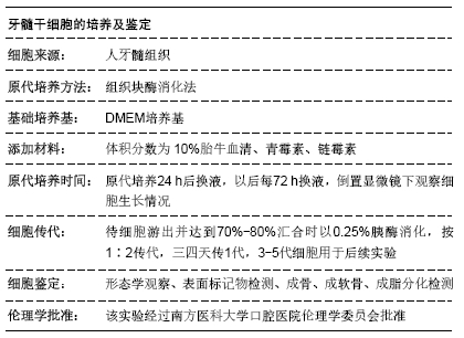
[5] 刘影,高杰,吴补领.改良组织块酶消化法原代培养人牙髓干细胞的研究[J].口腔疾病防治,2018,26(3):166-170.
[6] GRONTHOS S, MANKANI M, BRAHIM J, et al. Postnatal human dental pulp stem cells (DPSCs) in vitro and in vivo. Proc Natl Acad Sci U S A. 2000;97(25):13625-13630.
[7] ARTHUR A, RYCHKOV G, SHI S, et al. Adult human dental pulp stem cells differentiate toward functionally active neurons under appropriate environmental cues. Stem Cells. 2008;26(7):1787-1795.
[8] KIM JK, BAKER J, NOR JE, et al. mTor plays an important role in odontoblast differentiation. J Endod. 2011;37(8): 1081-1085.
[9] SUH JS, KIM KS, LEE JY, et al. A cell-permeable fusion protein for the mineralization of human dental pulp stem cells. J Dent Res. 2012;91(1):90-96.
[10] NAM S, WON JE, KIM CH, et al. Odontogenic differentiation of human dental pulp stem cells stimulated by the calcium phosphate porous granules. J Tissue Eng. 2011;2011:812547.
[11] GRONTHOS S, BRAHIM J, LI W, et al. Stem cell properties of human dental pulp stem cells. J Dent Res. 2002;81(8):531-535.
[12] WANG CY, TANI-ISHII N, STASHENKO P. Bone-resorptive cytokine gene expression in periapical lesions in the rat. Oral Microbiol Immunol. 1997;12(2):65-71.
[13] ZHANG X, NING T, WANG H, et al. Stathmin regulates the proliferation and odontoblastic/osteogenic differentiation of human dental pulp stem cells through Wnt/β-catenin signaling pathway. J Proteomics. 2019;202:103364.
[14] CHEN Y, GAO Y, TAO Y, et al. Identification of a Calcium-sensing Receptor in Human Dental Pulp Cells That Regulates Mineral Trioxide Aggregate-induced Mineralization. J Endod.2019;45(7):907-916.
[15] JIANG HW, ZHANG W, REN BP, et al. Expression of toll like receptor 4 in normal human odontoblasts and dental pulp tissue. J Endod. 2006;32(8):747-751.
[16] MUTOH N, TANI-ISHII N, TSUKINOKI K, et al. Expression of toll-like receptor 2 and 4 in dental pulp. J Endod. 2007;33(10): 1183-1186.
[17] ZHANG J, WU L, QU JM. Inhibited proliferation of human lung fibroblasts by LPS is through IL-6 and IL-8 release. Cytokine. 2011;54(3):289-295.
[18] SARRAZY V, VEDRENNE N, BILLET F, et al. TLR4 signal transduction pathways neutralize the effect of Fas signals on glioblastoma cell proliferation and migration. Cancer Lett. 2011;311(2):195-202.
[19] WANG YY, SUN SP, ZHU HS, et al. GABA regulates the proliferation and apoptosis of MAC-T cells through the LPS-induced TLR4 signaling pathway. Res Vet Sci. 2018; 118:395-402.
[20] YU L, ZHAO Y, GU X, et al. Dual effect of LPS on murine myeloid leukemia cells: Pro-proliferation and anti-proliferation. Exp Cell Res. 2016;344(2):210-218.
[21] PARK YD, KIM YS, JUNG YM, et al. Porphyromonas gingivalis lipopolysaccharide regulates interleukin (IL)-17 and IL-23 expression via SIRT1 modulation in human periodontal ligament cells. Cytokine. 2012;60(1):284-293.
[22] LI K, LV G, PAN L. Sirt1 alleviates LPS induced inflammation of periodontal ligament fibroblasts via downregulation of TLR4. Int J Biol Macromol. 2018;119:249-254.
[23] BOTERO TM, SHELBURNE CE, HOLLAND GR, et al. TLR4 mediates LPS-induced VEGF expression in odontoblasts. J Endod. 2006;32(10):951-955.
[24] NAKAO J, FUJII Y, KUSUYAMA J, et al. Low-intensity pulsed ultrasound (LIPUS) inhibits LPS-induced inflammatory responses of osteoblasts through TLR4-MyD88 dissociation. Bone. 2014;58:17-25.
[25] 刘影,高杰,吴补领.脂多糖与变形链球菌刺激DPSCs中Toll样受体4的表达[J].中国组织工程研究,2018,22(33):5333-5337.
[26] COIL J, TAM E, WATERFIELD JD. Proinflammatory cytokine profiles in pulp fibroblasts stimulated with lipopolysaccharide and methyl mercaptan. J Endod. 2004;30(2):88-91.
[27] NAGAOKA S, TOKUDA M, SAKUTA T, et al. Interleukin-8 gene expression by human dental pulp fibroblast in cultures stimulated with Prevotella intermedia lipopolysaccharide. J Endod.1996;22(1):9-12.
[28] TOKUDA M, SAKUTA T, FUSHUKU A, et al. Regulation of interleukin-6 expression in human dental pulp cell cultures stimulated with Prevotella intermedia lipopolysaccharide. J Endod. 2001;27(4):273-277.
[29] ADACHI T, NAKANISHI T, YUMOTO H, et al. Caries-related bacteria and cytokines induce CXCL10 in dental pulp. J Dent Res. 2007;86(12):1217-1222.
[30] PARK SH, HSIAO GY, HUANG GT. Role of substance P and calcitonin gene-related peptide in the regulation of interleukin-8 and monocyte chemotactic protein-1 expression in human dental pulp. Int Endod J. 2004;37(3):185-192.
[31] SILVA AC, FARIA MR, FONTES A, et al. Interleukin-1 beta and interleukin-8 in healthy and inflamed dental pulps. J Appl Oral Sci. 2009;17(5):527-532.
[32] LIU J, XU D, WANG Q, et al. LPS induced miR-181a promotes pancreatic cancer cell migration via targeting PTEN and MAP2K4. Dig Dis Sci. 2014;59(7):1452-1460.
[33] CHEN HJ, LIANG TM, LEE IJ, et al. Scutellariae radix suppresses LPS-induced liver endothelial cell activation and inhibits hepatic stellate cell migration. J Ethnopharmacol. 2013;150(3):835-842.
[34] HORTELANO S, LÓPEZ-FONTAL R, TRAVÉS PG, et al. ILK mediates LPS-induced vascular adhesion receptor expression and subsequent leucocyte trans-endothelial migration. Cardiovasc Res. 2010;86(2):283-292.
[35] LIU J, XU D, WANG Q, et al. LPS induced miR-181a promotes pancreatic cancer cell migration via targeting PTEN and MAP2K4. Dig Dis Sci. 2014;59(7):1452-1460.
[36] PETYAEV IM, ZIGANGIROVA NA, KOBETS NV, et al. Roquefort cheese proteins inhibit Chlamydia pneumoniae propagation and LPS-induced leukocyte migration. Scientific World Journal. 2013;2013:140591.
[37] WANG N, MENG X, LIU Y, et al. LPS promote Osteosarcoma invasion and migration through TLR4/HOTAIR.Gene. 2019; 680:1-8.
[38] LI D, FU L, ZHANG Y, et al. The effects of LPS on adhesion and migration of human dental pulp stem cells in vitro. J Dent. 2014;42(10):1327-1334.
[39] PARK JH, KWON SM, YOON HE, et al. Lipopolysaccharide promotes adhesion and migration of murine dental papilla-derived MDPC-23 cells via TLR4. Int J Mol Med. 2011; 27(2):277-281.
[40] 刘影,高杰,吴补领. TLR4 在人健康和深龋牙髓组织中的定位表达研究[J].口腔疾病防治,2017,25(3):153-158.
[41] MORSCZECK CO, DREES J, GOSAU M. Lipopolysaccharide from Escherichia coli but not from Porphyromonas gingivalis induce pro-inflammatory cytokines and alkaline phosphatase in dental follicle cells. Arch Oral Biol. 2012;57(12):1595-1601.
[42] GUO C, YUAN L, WANG JG, et al. Lipopolysaccharide (LPS) induces the apoptosis and inhibits osteoblast differentiation through JNK pathway in MC3T3-E1 cells. Inflammation. 2014; 37(2):621-631.
[43] PEI J, FAN L, NAN K, et al. Excessive Activation of TLR4/NF-κB Interactively Suppresses the Canonical Wnt/β-catenin Pathway and Induces SANFH in SD Rats. Sci Rep. 2017;7(1):11928.
[44] YU H, ZHANG X, SONG W, et al. Effects of 3-dimensional Bioprinting Alginate/Gelatin Hydrogel Scaffold Extract on Proliferation and Differentiation of Human Dental Pulp Stem Cells. J Endod. 2019;45(6):706-715.
|