| [1] Wu Y, Niu Y, Zhong S, et al. A preliminary investigation of the impact of oily skin on quality of life and concordance of self-perceived skin oiliness and skin surface lipids. Int J Cosmet Sci.2013;35(5): 442-447.[2] Iwona Driskell, Feride Oeztuerk-Winder, Peter Humphreys, et al. Genetically Induced Cell Death in Bulge Stem Cells Reveals Their Redundancy for Hair and Epidermal Regeneration. Stem Cells.2015;33(3): 988-998.[3] Marfia G, Navone SE, Di Vito C. Mesenchymal stem cells: potential for therapy and treatment of chronic non-healing skin wounds.Organogenesis.2015;11(4):183-206.[4] Hocking AM. The Role of chemokines in mesenchymal stem cell homing to wounds. Adv Wound Care (NewRochelle).2015;4(11): 623-630.[5] Wang Y, Liu ZY, Zhao Q, et al. Future application of hair follicle stem cells:capable in differentiation into sweat gland cells. Chin Med J (Engl).2013;126 (18): 3545-3552.[6] Purba TS, Haslam IS, Poblet E, et al. Human epithelial hair follicle stem cells and their progeny: current state of knowledge, the widening gap in translational research and future challenges. Bioessays.2014;36 (5): 513-525.[7] Mokos ZB,Mosler EL.Advances in a rapidly emerging field of hair follicle stem cell research. Collegium antropologicum.2014;38 (1): 373-378.[8] Kamachi Y,Kondoh H.Sox proteins: regulators of cell fate specification and differentiation. Development.2013;140(20): 4129-4144.[9] Gao Z, Cox JL, Gilmore JM, et al.Determination of Protein Interactome of Transcription Factor Sox2 in Embryonic Stem Cells Engineered for Inducible Expression of Four Reprogramming Factors.Angie Rizzino J Biol Chem.2012; 287(14):11384-11397.[10] Lien W H,Polak L,Lin M,et al.In vivo transcriptional governance of hair follicle stem cells by canonical Wnt regulators. Nat Cell Biol. 2014;16(2): 179 -190.[11] Rahmani W, Abbasi S, Hagner A, et al. Hair follicle dermal stem cells regenerate the dermal sheath,repopulate the dermal papilla,and modulate hair type. Dev Cell.2014;31(5): 543-558.[12] Rompolas P, Mesa KR, Greco V. Spatial organization within a niche as a determinant of stem-cell fate. Nature.2013;502(7472): 513-518.[13] Leiros GJ, Kusinsky AG, Drago H, et al. Dermal Papilla Cells Improve the Wound Healing Process and Generate Hair Bud-Like Structures in Grafted Skin Substitutes Using Hair Follicle Stem Cells. Stem Cells Transl Med.2014;3(10):1209-1219.[14] Najafzadeh N, Sagha M, Heydari Tajaddod S, et al. In vitro neural differentiation of CD34 (+) stem cell populations in hair follicles by three different neural induction protocols. Cell Dev Biol Anim.2015; 51(2): 192-203.[15] Dunn C,Wiltshire C,MacLaren A,et al.Molecular mechanism and biological functions of c-Jun N-terminal kinase signalling via the c-Jun transcription factor. Cell Signal.2002;14(7): 585-593.[16] Norifumi T, Rajan J, Matthew RL, et al. Hopx expression defines a subset of multipotent hair follicle stem cells and a progenitor population primed to give rise to K6+niche cells. Development. 2013;140(8):1655-1664.[17] Favaro R, Appolloni I, Pellegatta S, et al.Sox2 is required to maintain cancer stem cells in a mouse model of high-grade oligodendro-glioma. Cancer Res.2014;74:1833-1844.[18] Chen PL, Chen WS, Li J, et al.Diagnostic utlity of neural stem and progenitor cell markers nestin and Sox2 in distinguishing nodal melanocytic nevi from metastatic melanomas. Mod Pathol.2013; 26(1):44-53.[19] Jeong-Hyeon Lee, Won-Jae Lee, Ryoung-Hoon Jeon, et al. Development and Gene Expression of Porcine Cloned Embryos Derived from Bone Marrow Stem Cells with Overexpressing Oct4 and Sox2. Cell Reprogram.2014;16(6): 428-438.[20] Hsu YC, Li L, Fuchs E. Emerging interactions between skin stem cells and their niches[J]. Nat Med.2014;20(8):847-856.[21] Lee JH,Koh H,Kim M,et al. Energy-dependent regulation of cell structure by AMP-activated protein kinase. Nature.2007; 447 (7147):1017-1020.[22] Kuwano M, Horibe Y, Kawashima Y. Effect of collagen cross-linking in collagen corneal shields on ocular drug de-Livery. J Ocul Pharmacol Ther.1997;13(1):31-40.[23] Yamada N, Uchinuma E, Kuroyanagi Y. Clinical evaluation of an allogeneic cultured dermal substitute composed of fibroblasts within a spongy collagen matrix. Scand J Plast Reconstr Surg Hand Surg.1999;33(2):147-154.[24] Das A, Sinha M, Datta S, et al. Monocyte and macrophage plasticity in tissue repair and regeneration. Am J Pathol. 2015; 23(6):235-243.[25] Biswas SK, Chittezhath M, Shalova IN, et al. Macrophage polarization and plasticity in health and disease. Immunol Res. 2012;53(1/2/3):11-24.[26] Lo DD, Zimmermann AS, Nauta A, et al. Scarless fetal skin wound healing update. Birth Defects Res C Embryo Today, 2012, 96(30): 237-247.[27] Akita S, Akino K, Hirano A. Basic fi broblast growth factor in scarless wound healing. Adv Wound Care (New Rochelle). 2013; 2(2):44-49.[28] Green SA, Simoes CM, Bronner ME. Evolution of vertebrates as viewed from the crest. Nature. 2015;520(7548): 474-482.[29] Mozafari S, Laterza C, Roussel D, et al. Skin- derived neural precursors competitively generate functional myelin in adult demyelinated mice. J Clin Invest.2015;125(9): 3642-3656.[30] Sato H, Ebisawa K, Takanari K, et al. Skin-derived precursor cells promote wound healing in diabetic mice.Ann Plast Surg.2015; 74(1):114-120.[31] Ochalek M, Hleiss RS, Wohl AB J, et al. Characterization of lipid model membranes designed for studying impact of ceramide species on drug diffusion and penetration.Eur J Pharm Biopharm. 2012;81(1):113-120.[32] Rahmani W, Abbasi S, Hagner A, et al. Hair follicle dermal stem cells regenerate the dermal sheath, repopulate the dermal papilla,and modulate hair type. Dev Cell.2014;31(5): 543-558.[33] Shu B, Xie JL, Xu YB, et al. Directed differentiation of skin-derived precursors into fibroblast-like cells. Int J Clin Exp Pathol.2014; 7(4):1478-1486.[34] Mehrabi M,Mansouri K,Hosseinkhani S,et al.Differentiation of human skin-derived precursor cells into functional islet-like insulin-producing cell clusters. In Vitro Cell Dev Biol Anim.2015;51 (6):595-603.[35] Thangapazham RL,Darling TN,Meyerle J.Alteration of skin properties with autologous dermal fibroblasts.Int J Molecul Sci. 2014;15(5):8407-8427.[36] Huang Z, Zhen Y, Yin W, Ma Z, Zhang L. Shh promotes sweat gland cell maturation in three-dimensional culture. Cell Tissue Banking.2016;17(2):317-325.[37] Boekema B,Boekestijn B,Breederveld RS.Evaluation of saline,RPMI and DMEM/F12 for storage of split-thickness skin grafts.Burns.2015;41(4):848-852.[38] Zhang C, Chen Y, Fu X. Sweat gland regeneration after burn injury: Is stem cell therapy a new hope. Cytotherapy.2015; 17(5):526-535.[39] Zhao Z, Xu M, Wu M, et al. Direct reprogramming of human fibroblasts into sweat gland-like cells. Cell Cycle.2015;14(21): 3498-3505.[40] Joannides A, Gaughwin P, Schwiening C, et al. Efficient generation of neural precursors from adult human skin:astrocytes promote neurogenesis from skin-derived stem cells. Lancet. 2004; 364(9429):172-178.[41] Ikeda R, Ling J, Cha M, et al. In situ patch-clamp recordings from Merkel cells in rat whisker hair follicles, an experimental protocol for studying tactile transduction in tactile-end organs. Mol Pain. 2015;11(23):15-22.[42] Bruns I,Lucas D,Pinho S,et al.Megakaryocytes regulate hematopoietic stem cell quiescence through CXCL4 secretion . Nature Med.2014;20(11):1315-1320.[43] Andersen DC,Skovrind I,Christensen ML, et al. Stem cell survival is severely compromised by the thymidine analog EdU, an alternative to BrdU for proliferation assays and stem cell tracing. Anal Bioanal Chem.2013;405(29):9585-9591. |
.jpg) 文题释义:
毛囊:在真皮乳头层里,毛根被内、外毛鞘包围,外毛鞘又被结缔组织细胞包围,形成口袋状,称毛囊。毛囊是包围在毛发根部的囊状组织,内层是上皮组织性毛囊,外层是结缔组织性毛囊,内层与表皮相连,外层则与真皮相连。毛囊由毛杆、毛凸、毛球、毛乳头、毛基质等部分组成。神经纤维末梢使毛囊具有感觉的功能,动脉和静脉毛细血管丛给毛囊提供血液。毛囊的最深处是位于角质层3-7 mm的毛乳头,它含有神经和血管,向毛杆提供养分。毛囊的发育经过表皮和真皮之间一系列复杂的相互作用而形成,而具有周期性。
皮肤源性前体细胞:是一种神经嵴来源的前体细胞,具有来源丰富、易于获取和多向分化的潜能等优点,能够分化为在体内外都有功能的许旺细胞、骨样细胞和胰岛素样细胞等多种细胞类型。其在多种组织修复和疾病治疗,特别是神经损伤再修复中显示了良好的应用前景。
文题释义:
毛囊:在真皮乳头层里,毛根被内、外毛鞘包围,外毛鞘又被结缔组织细胞包围,形成口袋状,称毛囊。毛囊是包围在毛发根部的囊状组织,内层是上皮组织性毛囊,外层是结缔组织性毛囊,内层与表皮相连,外层则与真皮相连。毛囊由毛杆、毛凸、毛球、毛乳头、毛基质等部分组成。神经纤维末梢使毛囊具有感觉的功能,动脉和静脉毛细血管丛给毛囊提供血液。毛囊的最深处是位于角质层3-7 mm的毛乳头,它含有神经和血管,向毛杆提供养分。毛囊的发育经过表皮和真皮之间一系列复杂的相互作用而形成,而具有周期性。
皮肤源性前体细胞:是一种神经嵴来源的前体细胞,具有来源丰富、易于获取和多向分化的潜能等优点,能够分化为在体内外都有功能的许旺细胞、骨样细胞和胰岛素样细胞等多种细胞类型。其在多种组织修复和疾病治疗,特别是神经损伤再修复中显示了良好的应用前景。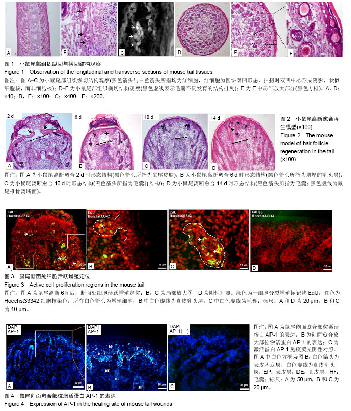