中国组织工程研究 ›› 2015, Vol. 19 ›› Issue (23): 3736-3743.doi: 10.3969/j.issn.2095-4344.2015.23.022

• 骨髓干细胞 bone marrow stem cells • 上一篇    下一篇

骨髓间充质干细胞诱导胆管内皮细胞与电纺纳米纤维的生物相容性

杨  扬1,周家华1,殷雪琰2,徐  勇1,曹  阳2,许  茜2   

  1. 1东南大学附属中大医院肝胆胰中心,江苏省南京市  210009; 
    2东南大学公共卫生学院,江苏省南京市  210009
  • 出版日期:2015-06-04 发布日期:2015-06-04
  • 通讯作者: 周家华,博士,教授,主任医师,东南大学附属中大医院肝胆胰中心,江苏省南京市 210009
  • 作者简介:杨扬,男,1985年生,山西省盂县人,汉族,2011年东南大学毕业,硕士,医师,主要从事胰腺与胆道疾病的临床与基础研究。
  • 基金资助:

    江苏省卫生厅医学基金项目(H200815),项目名称:组织工程胆管的应用研究

Biocompatibility of porcine bone marrow mesenchymal stem cells-derived bile duct endothelial cells with electrospun nanofibers

Yang Yang1, Zhou Jia-hua1, Yin Xue-yan2, Xu Yong1, Cao Yang2, Xu Qian2   

  1. 1Hepatobiliary & Pancreatic Center, Zhongda Hospital, Southeast University, Nanjing 210009, Jiangsu Province, China; 
    2College of Public Health, Southeast University, Nanjing 210009, Jiangsu Province, China
  • Online:2015-06-04 Published:2015-06-04
  • Contact: Zhou Jia-hua, M.D., Professor, Chief physician, Hepatobiliary & Pancreatic Center, Zhongda Hospital, Southeast University, Nanjing 210009, Jiangsu Province, China
  • About author:Yang Yang, Master, Physician, Hepatobiliary & Pancreatic Center, Zhongda Hospital, Southeast University, Nanjing 210009, Jiangsu Province, China
  • Supported by:

    Medical Foundation of Department of Public Health of Jiangsu Province of China, No. H200815

摘要:

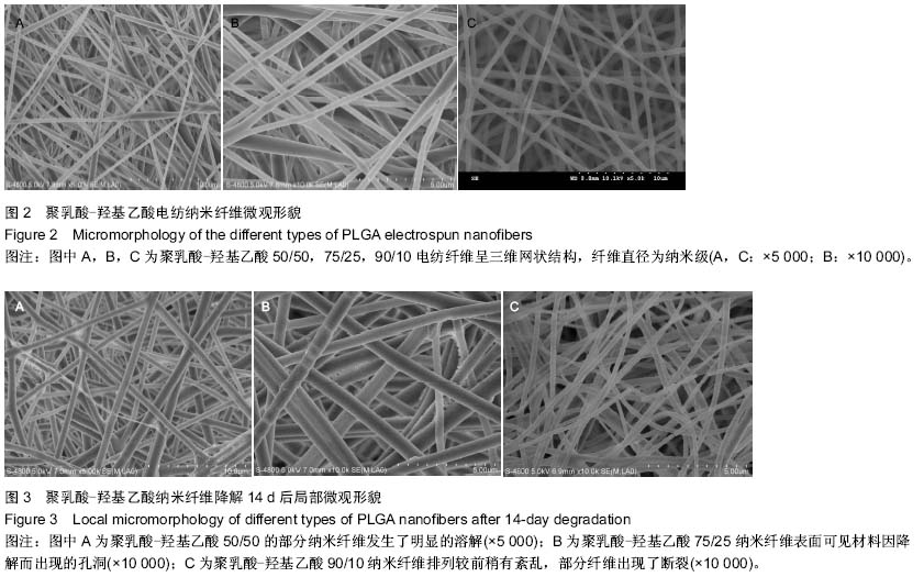
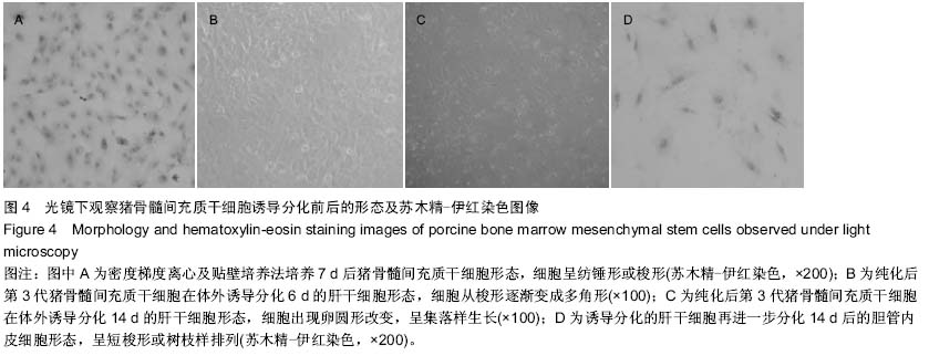
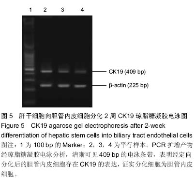
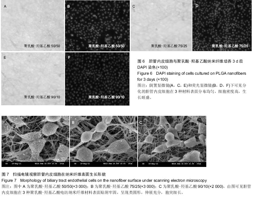
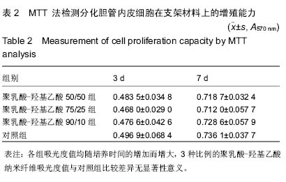
背景:胆管损伤修复是腹部外科手术的难题,组织工程胆管是解决这一难题的理想方法,而构建性能优异的组织工程胆管支架是这一研究的关键。
目的:观察猪骨髓间充质干细胞诱导分化的胆管内皮细胞与电纺纳米纤维的生物相容性。
方法:将猪骨髓间充质干细胞于体外定向分化为肝干细胞,再进一步分化为胆管内皮细胞,并通过形态学及RT-PCR对分化完成的细胞加以鉴定。用静电纺丝方法制备聚乳酸-羟基乙酸纳米纤维膜,通过扫描电镜观察其形态,并测定其短期(2周)内体外降解率。将分化的胆管内皮细胞与纳米纤维复合培养,观察细胞在纳米纤维表面的黏附、增殖能力,荧光染色观察细胞形态及在材料表面的分布情况,扫描电镜观察细胞在纳米纤维表面生长形貌。
结果与结论:骨髓间充质干细胞体外定向分化4周后,细胞呈现典型树枝状胆管内皮细胞形态学改变,并且有CK19的表达。扫描电镜照片显示电纺材料为连续的纳米纤维,纤维直径均分布在200-500 nm范围内,在2周内聚乳酸-羟基乙酸纳米纤维没有发生明显的降解。通过细胞黏附率计算、MTT法检测、荧光染色及扫描电镜观察证明猪骨髓间充质干细胞诱导分化后的胆管内皮细胞与聚乳酸-羟基乙酸纳米纤维具有良好的生物相容性。

中国组织工程研究杂志出版内容重点:干细胞;骨髓干细胞;造血干细胞;脂肪干细胞;肿瘤干细胞;胚胎干细胞;脐带脐血干细胞;干细胞诱导;干细胞分化;组织工程

关键词: 干细胞, 分化, 组织工程胆管, 骨髓间充质干细胞, 胆管内皮细胞, 电纺纳米纤维

Abstract:

BACKGROUND: Repair of extrahepatic biliary tract injury is a difficult problem in the abdominal surgery. Tissue-engineered extrahepatic biliary tract is an ideal selection for this problem. Construction of tissue-engineered extrahepatic biliary tract with excellent performance is a key to related studies.
OBJECTIVE: To investigate the biocompatibility of bile duct endothelial cells differentiated by porcine bone marrow mesenchymal stem cells with electrospun nanofibers.
METHODS: Porcine bone marrow mesenchymal stem cells were induced toward biliary tract endothelial cells, which were then identified by morphology and RT-PCR. Polylactic-co-glycolic acid (PLGA) nanofiber membranes were prepared by electrospinning. The morphology was determined by scanning electron microscopy and the short-term (2-week) in vitro degradation rate was determined. Adhesion and proliferation of biliary tract endothelial cells on the nanofiber surface was analyzed by calculating the cell adhesion rate and MTT assay, respectively. Cell growth, morphology and distribution on the material surface were observed by fluorescence staining and scanning electron microscopy, respectively.
RESULTS AND CONCLUSION: After 4 weeks of directed differentiation of bone marrow mesenchymal stem cells in vitro, cells showed typical morphology of dendritic bile duct endothelial cells and had the expression of CK19. Scanning electron micrographs showed that electrospun materials were continuous nanofibers with diameters between 200 and 500 nm. No significant degradation of the PLGA nanofibers was observed within 2 weeks. Based on the measured cell adhesion rate, MTT assay, fluorescence staining, and scanning electron microscopy, the differentiated cells possessed a good proliferative capacity on PLGA nanofibers. Bone marrow mesenchymal stem cells differentiated into bile duct endothelial cells in vitro. Materials prepared by the electrospinning method had a nanofiber structure, which did not significantly degrade within 2 weeks. Differentiated cells exhibit good biocompatibility with the nanofibers.

中国组织工程研究杂志出版内容重点:干细胞;骨髓干细胞;造血干细胞;脂肪干细胞;肿瘤干细胞;胚胎干细胞;脐带脐血干细胞;干细胞诱导;干细胞分化;组织工程

Key words: Bone Marrow, Mesenchymal Stem Cells, Cell Differentiation, Bile Duct Endothelial Cells, Nanofibers, Biocompatible Materials

中图分类号: