中国组织工程研究 ›› 2015, Vol. 19 ›› Issue (23): 3711-3715.doi: 10.3969/j.issn.2095-4344.2015.23.018

• 干细胞移植 stem cell transplantation • 上一篇    下一篇

经气管移植骨髓间充质干细胞在染矽尘大鼠体内的归巢

黄  明1,周永梅1,李  斌1,吴奇峰1,朱玉峰2,梁伟辉1   

  1. 1广东省职业病防治院,广东省广州市  510300; 
    2南方医科大学南方医院实验动物研究中心,广东省广州市  510515
  • 出版日期:2015-06-04 发布日期:2015-06-04
  • 作者简介:黄明,女,1980年生,湖南省平江县人,汉族,2010年南方医科大学毕业,博士,副主任医师,主要从事职业病防治研究。
  • 基金资助:

    国家自然科学基金项目(81302396);国家十二五科技支撑计划分课题(2014BAI12B01);广东省职业病防治重点实验室(2012A 061400007)

The homing of bone marrow mesenchymal stem cells transplanted via the trachea into rats exposed to silica dust 

Huang Ming1, Zhou Yong-mei2, Li Bin1, Wu Qi-feng1, Zhu Yu-feng2, Liang Wei-hui1   

  1. 1Guangdong Province Hospital for Occupational Disease Prevention and Treatment, Guangzhou 510300, Guangdong Province, China; 
    2Center for Experimental Animals, Nanfang Hospital, Southern Medical University, Guangzhou 510515, Guangdong Province, China
  • Online:2015-06-04 Published:2015-06-04
  • About author:Huang Ming, M.D., Associate chief physician, Guangdong Province Hospital for Occupational Disease Prevention and Treatment, Guangzhou 510300, Guangdong Province, China
  • Supported by:

    the National Natural Science Foundation of China, No. 81302396; the National Science and Technology Support Plan during the 12th Five-Year Period, No. 2014BAI12B01; Guangdong Key Laboratory for Occupational Disease Prevention, No. 2012A061400007

摘要:

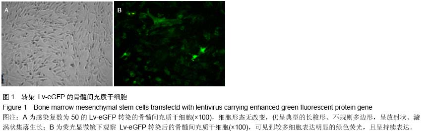
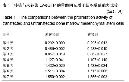
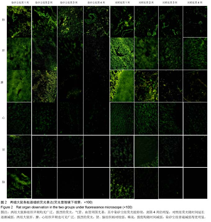
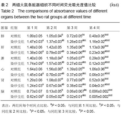
背景:骨髓间充质干细胞经气管途径注入大鼠体内能减轻染矽尘大鼠的肺部损伤,但其在大鼠体内的分布、迁移情况尚不清楚。
目的:观察经气管途径移植的骨髓间充质干细胞在染矽尘大鼠体内的分布和归巢情况。
方法:用携带增强型绿色荧光蛋白基因的慢病毒(Lv-eGFP)转染SD大鼠骨髓间充质干细胞,锥虫蓝染色和CCK-8法检测转染前后细胞的活力及增殖情况。将SPF级SD大鼠随机分为染矽尘组和对照组,染矽尘组大鼠经气管内注入40 g/L无菌矽尘混悬液1 mL,对照组大鼠注入生理盐水1 mL。造模后第2天每只大鼠经气管注入2.5×10⁶个Lv-eGFP转染的骨髓间充质干细胞,分别于移植后的1,2,3,4周处死大鼠,取心、肝、脾、肺、肾、脑组织冰冻切片,荧光显微镜观察各组织荧光表达,图文分析软件计算组织荧光强度。  
结果与结论:感染复数为50时,转染与未转染Lv-eGFP的骨髓间充质干细胞在活细胞率及增殖力上差异无显著性意义(P > 0.05)。注入细胞后两组肺组织切片均可见广泛、强烈的绿色荧光,以支气管、血管周围明显,各时间点染矽尘组的荧光强度均高于对照组(P < 0.05),到第4周荧光仍较强。两组其余各脏器在第1周均可见荧光,肝、脾、心脏荧光强、分布广,肾、脑组织荧光相对较弱、分布较少;随时间推移,荧光均逐渐减弱,分布逐渐减少。结果提示骨髓间充质干细胞经气管途径进入染矽尘大鼠体内后可归巢至受损肺部。

中国组织工程研究杂志出版内容重点:干细胞;骨髓干细胞;造血干细胞;脂肪干细胞;肿瘤干细胞;胚胎干细胞;脐带脐血干细胞;干细胞诱导;干细胞分化;组织工程

关键词: 干细胞, 移植, 骨髓间充质干细胞, 绿色荧光蛋白, 二氧化硅, 移植, 气管途径, 归巢, 国家自然科学基金

Abstract:

BACKGROUND: Bone marrow mesenchymal stem cells transplantation via the trachea can relieve the lung injury of rats exposed to silica dust, but their distribution and migration in vivo is still unclear.
OBJECTIVE: To investigate the distribution and homing of bone marrow mesenchymal stem cells transplanted via the trachea into rats exposed to silica dust.
METHODS: Bone marrow mesenchymal stem cells from Sprague-Dawley rats were isolated through bone marrow adherent method and transfected with lentivirus carrying enhanced green fluorescent protein gene (Lv-eGFP). Trypan blue staining and cell counting kit-8 were applied to assay the viability and proliferation of the transfected and untransfected cells. Sprague-Dawley rats, SPF level, were randomized into control group and silica dust exposure group. Rats in the two groups were respectively injected via the trachea with 1 mL of sterile 
silica dust suspensions (40 g/L) and 1 mL of normal saline. At 2 days after modeling, 2.2×10⁶ transfected bone marrow mesenchymal stem cells were injected via the trachea into the rats of control group and silica dust exposure group. Rats were killed at weeks 1, 2, 3, 4 after transplantation, and the distribution and intensity of green fluorescence in the lung, heart, liver, spleen, kidney, and brain tissue were observed under the fluorescence microscopy by frozen sections and analyzed using imaging analysis software.
RESULTS AND CONCLUSION: When the multiplicity of infection was 50, there were no significant differences between the viability and proliferation activity of the transfected and untransfected cells (P > 0.05). After transplantation of transfected bone marrow mesenchymal stem cells, strong green fluorescence was observed widely in the lung, especially around the bronchus and blood vessels, and still obvious at the 4th week. The fluorescence of other organs also could be observed at the 1st week. It was strong and wide in the liver, spleen and heart, while faint and less in the kidney and brain, and all reduced with time. It shows bone marrow mesenchymal stem cells transplanted via the trachea into rats exposed to silica dust can be homing to the injured lung of rats.

中国组织工程研究杂志出版内容重点:干细胞;骨髓干细胞;造血干细胞;脂肪干细胞;肿瘤干细胞;胚胎干细胞;脐带脐血干细胞;干细胞诱导;干细胞分化;组织工程

Key words: Bone Marrow, Mesenchymal Stem Cell Transplantation, Lung, Drug Administration Routes

中图分类号: