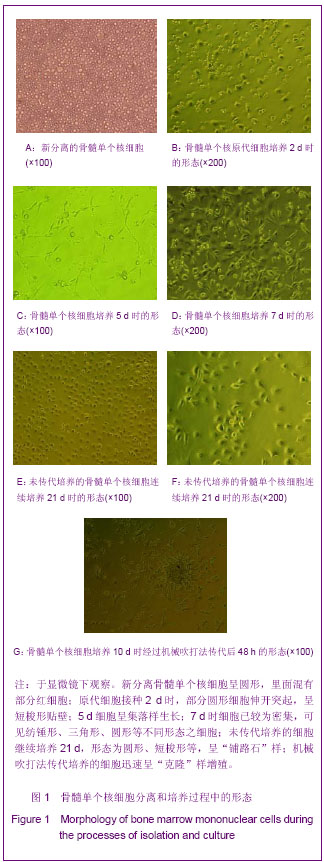
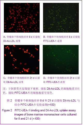
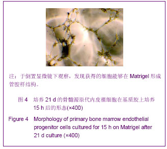
| [1] Hur J, Yoon CH, Kim HS, et al. Characterization of two types of endothelial progenitor cells and their different contributions to neovasculogenesis. Arterioscler Thromb Vasc Biol. 2004; 24(2):288-293.[2] Dzau VJ, Gnecchi M, Pachori AS, et al. Therapeutic potential of endothelial progenitor cells in cardiovascular diseases. Hypertension. 2005;46(1):7-18.[3] Urbich C, Dimmeler S. Endothelial progenitor cells: Characterization and role in vascular biology. Circ Res. 2004; 95(4):343-353.[4] Asahara T, Mumhara T, Suilivan A, et al. Isolation of Putative Progenitor endothelial cells for angiogenesis. Science. 1997; 275 (5302):964-967.[5] Asahara T, Masuda H, Takahashi T, et al. Bone Marrow Origin of Endothelial Progenitor Cells Responsible for Postnatal Vasculogenesis in Physiological and Pathological Neovascularization. Circ Res. 1999;85(3):221-228.[6] Morgan R, Kreipke CW, Roberts G, et al. Neovascularization following traumatic brain injury:possible evidence for both angiogenesis and vasculogenesis. Neurol Res. 2007;29(4): 375-381.[7] Umemura T, Higashi Y. Endothelial progenitor cells:therapeutic target for cardiovascular diseases. J Pharmacol Sci. 2008;108(1):1-6.[8] Gao D, Nolan DJ, Mellick AS, et al. Endothelial progenitor cells control the angiogenic switch in mouse lung metastasis. Science. 2008;319(5860):195-198.[9] Su Y, Zheng L, Wang Q, et al. The PI3K/Akt pathway upregulates Id1 and integrin α4 to enhance recruitment of human ovarian cancer endothelial progenitor cells. BMC Cancer. 2010;10:459.[10] Moubarik C, Guillet B, Youssef B, et al. Transplanted late outgrowth endothelial progenitor cells as cell therapy product for stroke. Stem cell reviews. 2011;7(1):208-220.[11] Rouwkema J, Westerweel PE, de Boer J, et al. The use of endothelial progenitor cells for prevascularized bone tissue engineering.Tssue Eng Part A. 2009;15(8):2015-2027.[12] Asahara T. Cell therapy and gene therapy using endothelial progenitor cells for vascular regeneration. Handb Exp Pharmacol. 2007;(180):181-194.[13] Palladino M, Gatto l, Neri V, et al. Combined Therapy with Sonic Hedgehog Gene Transfer and Bone Marrow-Derived Endothelial Progenitor Cells Enhances Angiogenesis and Myogenesis in the Ischemic Skeletal Muscle. J Vasc Res. 2012;49(5):425-431.[14] Weber A, Pedrosa I, Kawamoto A, et al. Magnetic resonance mapping of transplanted endothelial progenitor cells for therapeutic neovascularization in ischemic heart disease. Eur J Cardiothorac Surg. 2004;26(1):137-143.[15] Khan SS, Solomon MA, McCoy JP Jr. Detection of circulating endothelial cells and endothelial progenitor cells by flow cytometry. Cytometry B Clin Cytom. 2005;64(1):l-8.[16] Medina RJ, O'Neill CL, Sweeney M, et al. Molecular analysis of endothelial progenitor cell (EPC) subtypes reveals two distinct cell populations with different identities. BMC Med Genomics. 2010;3:18.[17] Yuan JM, Guan SH, Dang RS, et al.Zhongguo Zuzhi Gongcheng Yanjiu yu Linchuang Kangfu. 2010;14(36): 6662-6666.袁建明,管松晖,党瑞山,等.绵羊骨髓来源内皮祖细胞的培养与鉴定[J].中国组织工程研究与临床康复,2010,14(36):6662-6666.[18] Asai J, Takenaka H, Li M, et al. Topical application of ex vivo expanded endothelial progenitor cells promotes vascularisation and wound healing in diabetic mice. Int Wound J.2012.[19] Zhao T, Li J, Chen AF. MicroRNA-34a induces endothelial progenitor cell senescence and impedes its angiogenesis via suppressing silent information regulator 1. Am J Physiol Endocrinol Metab. 2010;299(1):E110-E116.[20] Brunt KR, Hall SR, Ward CA, et al. Endothelial Progenitor Cell and Mesenchymal Stem Cell Isolation, Characterization, Viral Transduction. Methods Mol Med. 2007;139:197-210. [21] Chen J, Song M, Yu S, et at. Advanced glycation endproducts alter functions and promote apoptos in endothelial progenitor cells through receptor for advanced glycation endproducts mediate over pression of cell oxidant stress. Mol Cell Biochem. 2010;335(1-2):137-146.[22] li M. Bone Marrow-Derived Endothelial Progenitor Cells:Isolation and Characterization for Myocardial Repair. Methods Mol Biol. 2010;660:9-27.[23] Ahrens I, Domeij H, Topcic D, et al. Successful in vitro expansion an differentiation of cord blood derived CD34+ cells into early endothelial progenitor cells reveals highly differential gene expression. PLoS One. 2011; 6(8):e23210.[24] Thill M, Strunnikova NV, Berna MJ, et al. Late outgrowth endothelial progenitor cells in patients with age-related macular degeneration. Invest Ophthalmol Vis Sci. 2008; 49(6):2696-2708.[25] Umemura T, Higashi Y. Endothelial progenitor cells:therapeutic target for cardiovascular diseases. J Pharmacol Sci. 2008;108(1):1-6.[26] Garcia-Barros M, Paris F, Cordon-Cardo C, et al. Tumor Response to Radiotherapy Regulated by Endothelial Cell Apoptosis. Science. 2003;300(5622):1155-1159.[27] Ingram DA, Mead LE, Tanaka H, et al. Identification of a novel hierarchy of endothelial progenitor cells using human peripheral and umbilical cord blood. Blood. 2004;104(9): 2752-2760.[28] Werner N, Junk S, Laufs U, et al. Intravenous Transfusion of Endothelial Progenitor Cells Reduces Neointima Formation After Vascular Injury. Circ Res. 2003;93(2):e17-e24.[29] Cherqui S, Kurian SM, Schussler O, et al. Isolation and angiogenesis by endothelial progenitors in the fetal liver. Stem Cells. 2006;24(1):44-54.[30] Casamassimi A, Balestrieri ML, Fiorito C, et al. Comparison between total endothelial progenitor cell isolation versus enriched CD133+ culture. J Biochem. 2007;141(4):503-511.[31] Kahler CM, Wechselberger J, Hilbe W, et al. Peripheral infusion of rat bone marrow derived endothelial progenitor cells leads to homing in acute lung injury. Respir Res. 2007; 8(1):50.[32] Chen YH, Lin SJ, Lin F Y, et al. High glucose impairs early and late endothelial progenitor cells by modifying nitric oxide-related but not oxidative stress-mediated mechanism. Diabetes. 2007;56(6):1559-1568[33] Hirschi KK, Ingram DA, Yoder MC. Assessing identity,phenotype,and fate of endothelial progenitor cells. Arterioscle Thromb Vasc Biol. 2008;28(9):1584-1589.[34] Yoder MC. Defining human endothelial progenitor cells. J Thromb Haemost. 2009;7 Suppl 1:49-52.[35] Dimmeler S. Regulation of bone marrow-derived vascular progenitor cell mobilization and maintenance. Arterioscler Thromb Vasc Biol. 2010;30(6):1088-1093.[36] Leone AM, Valgimigli M, Giannico MB, et al. From bone marrow to the arterial wall:the ongoing tale of endothelial progenitor cells. Eur Heart. 2009;30(8):890-899.[37] Asahara T, Kawamoto A. Endothelial progenitor cells for postnatal vasculogenesis. Am J Physiol Cell Physiol. 2004; 287(3):C572-579.[38] Barsotti MC, Magera A, Armani C, et al. Fibrin acts as biomimetic niche inducing both differentiation and stem cell marker expression of early human endothelial progenitor cells. Cell Prolif. 2011;44(1):33-48.[39] Bleiziffer O, Hammon M, Naschberger E, et al. Endothelial progenitor cells are integrated in newly formed capillaries and alter adjacent fibrovascular tissue after subcutaneous implantation in a fibrin matrix. J Cell Mol Med. 2011;15: 2452-2461.[40] Shi S, He YZ, Song L, et al. Zhongguo Zuzhi Gongcheng Yanjiu yu Linchuang Kangfu. 2010;14(16):2879-2882. 施森,何延政,宋丽,等.血管内皮生长因子在鼠尾胶原凝胶诱导三维血管新生中的作用研究[J].中国组织工程研究与临床康复, 2010, 14(16):2879-2882. |

.jpg)