[1] 夏冬景.3种冠边缘肩台设计对龈沟液中IL-1β、AST及其OPN水平的影响[J].口腔医学研究,2013,29(12):1161-1164.
[2] 邢成岗,朱岩凤,李冠颖.不同口腔修复材料表面细菌黏附和生长情况的对比分析[J].全科口腔医学电子杂志,2015,2(8):59-60.
[3] 刘镇凡,梁晓.全瓷冠修复体对基牙龈沟液影响的临床研究[J].广西医科大学学报,2015,32(4):611-613.
[4] 王亚玲,曹直,占时霞.钴铬合金和金合金烤瓷全冠修复对龈沟液中AST、ALP、TNF-α和IL-8、GP-x、MDA水平的影响[J].海南医学院学报,2016,22(17):2069-2072.
[5] 马玉龙,王海山,那日苏.钴铬合金烤瓷冠和全瓷冠修复对患牙牙周组织及龈沟液中炎症因子水平的影响研究[J].中国实用口腔科杂志,2016,9(12):747-750.
[6] 郭斌,孙雷,翟晓红.氧化锆全瓷全冠修复对龈沟液患者的临床疗效观察[J].河北医学,2017,23(10):1632-1636.
[7] 王一立.不同烤瓷冠修复对牙周组织龈沟出血情况及血管内皮生长因子和肿瘤坏死因子α水平变化分析[J].中国临床医生杂志, 2017,45(9):89-91.
[8] 赵恒越,何文娟.三种修复金属材料对口腔修复患者牙周组织龈沟液中炎症因子水平的影响[J].河北医学,2019,25(6):894-898.
[9] LIU Y, ZHAO R, REDA B, et al. Profiling of cytokines, chemokines and growth factors in saliva and gingival crevicular fluid. Cytokine. 2021; 142:155504.
[10] 张彬,刘琨,李克义,等.3种全冠修复对患者焦虑抑郁心理状况及龈沟液中IL-17、IL-35水平的影响[A].中华口腔医学会全科口腔医学专业委员会、中国科学技术协会国际科技交流中心.中华口腔医学会全科口腔医学专业委员会第八次学术会议论文集[C].中华口腔医学会全科口腔医学专业委员会、中国科学技术协会国际科技交流中心:中华口腔医学会,2017:1.
[11] 李学盛,李鸿波.固定修复体适应性评价方法的研究进展[J].国际口腔医学杂志,2017,44(6):726-730.
[12] 孙培音,李会云,谭为聪,等.不同种类烤瓷肩台对基牙牙周组织的影响[J].全科口腔医学电子杂志,2015,2(2):84-85.
[13] NEMANE V, AKULWAR RS, MESHRAM S. The effect of various finish line configurations on the marginal seal and occlusal discrepancy of cast full crowns after cementation-an in-vitro study. J Clin Diagn Res. 2015; 9(8):ZC18-ZC21.
[14] 杨乐.全瓷冠龈上与平龈边缘位置的选择对牙龈的影响差异[J].医学美学美容,2018,27(19):65.
[15] 杨静.烤瓷熔附金属冠颈缘位置对牙周组织的影响[D].昆明:昆明医科大学,2016.
[16] KHURSHID Z, MALI M, NASEEM M, et al. Human gingival crevicular fluids (GCF) proteomics: an overview. dent J (Basel). 2017;5(1):12.
[17] KHURSHID Z, WARSI I, MOIN SF, et al. Biochemical analysis of oral fluids for disease detection. Adv Clin Chem. 2021;100:205-253.
[18] COSTANTINI E, SINJARI B, PISCOPO F, et al. Evaluation of salivary cytokines and vitamin d levels in periodontopathic patients. Int J Mol Sci. 2020;21(8):2669.
[19] 陈崇崇,钟良军.龈沟液生物标志物在慢性牙周炎诊疗中的研究进展[J].口腔医学,2019,39(11):1047-1052.
[20] 弓飞.氧化锆全瓷与钴铬合金全冠修复牙列、牙体缺损临床效果对比[J].实用中西医结合临床,2020,20(1):57-58.
[21] 陈珂,舒成军,陈梦铮.二氧化锆全瓷冠对前牙牙体缺损的临床疗效及对周围组织的影响[J].现代实用医学,2020,32(4):509-511.
[22] 胡竹林,赵诣,李茵.口腔龈沟液生物标志物的检测分析现状及临床应用前景展望[J].国际口腔医学杂志,2019,46(3):308-315.
[23] 林晓丽.不同表面处理对氧化锆表面粗糙度和细菌黏附影响的研究[D].福州:福建医科大学,2017.
[24] 李龙飞.不同修复材料对口腔修复患者炎性指标的影响比较[J].当代医学,2020,26(31):125-126.
[25] 杜花娇,刘族志,吴小芳.二氧化锆全瓷冠修复前牙缺损效果研究[J].创伤与急危重病医学,2020,8(1):56-57.
[26] 贾浩,宁静,王培,等.全瓷冠与金属烤瓷冠在前牙缺损修复中的疗效比较[J].河北医药,2018,40(12):1880-1883.
[27] HEBOYAN A, SYED AUY, ROKAYA D, et al. Cytomorphometric analysis of inflammation dynamics in the periodontium following the use of fixed dental prostheses. Molecules. 2020;25(20):4650.
[28] TU Y, LING X, CHEN Y, et al. Effect of S. Mutans and S. Sanguinis on Growth and Adhesion of P. Gingivalis and Their Ability to Adhere to Different Dental Materials. Med Sci Monit. 2017;23:4539-5445.
[29] 夏丽,储冰峰.变形链球菌LuxS蛋白与LuxS/AI-2群体感应系统的研究进展[J].中华老年口腔医学杂志,2011,9(5):303-306.
[30] 杜留熠,吕慧欣,王鹞,等.牙龈卟啉单胞菌fimA分型与相关疾病的研究[J].口腔医学研究,2018,34(12):1281-1283.
[31] BALASHOVA N, DHINGRA A, BOESZE-BATTAGLIA K, et al. Aggregatibacter actinomycetemcomitans leukotoxin induces cytosol acidification in LFA-1 expressing immune cells. Mol Oral Microbiol. 2016;31(1):106-114.
[32] FATIMA T, KHURSHID Z, REHMAN A, et al. Gingival Crevicular Fluid (GCF): A Diagnostic Tool for the Detection of Periodontal Health and Diseases. Molecules. 2021;26(5):1208.
[33] SHOJI M, TAKESHITA T, MARUYAMA F, et al. Recent advances in the field of oral bacteriology. Nihon Saikingaku Zasshi. 2015;70(2):333-338.
[34] MATHEW MG, SAMUEL SR, SONI AJ, et al. Evaluation of adhesion of Streptococcus mutans, plaque accumulation on zirconia and stainless steel crowns, and surrounding gingival inflammation in primary molars: randomized controlled trial. Clin Oral Investig. 2020;24(9):3275-3280.
[35] DOBRZYNSKI M, PAJACZKOWSKA M, NOWICKA J, et al. Study of Surface Structure Changes for Selected Ceramics Used in the CAD/CAM System on the Degree of Microbial Colonization, In Vitro Tests. Biomed Res Int. 2019;2019:9130806.
[36] HAO Y, HUANG X, ZHOU X, et al. Influence of Dental Prosthesis and Restorative Materials Interface on Oral Biofilms. Int J Mol Sci. 2018; 19(10):3157.
[37] 高茜.不同工艺制作的钴铬合金和钛的细菌黏附及耐腐蚀性研究[D].济南:山东大学,2020.
[38] HEBOYAN A, MANRIKYAN M, ZAFAR MS, et al. Bacteriological evaluation of gingival crevicular fluid in teeth restored using fixed dental prostheses: an In vivo study. Int J Mol Sci. 2021;22(11):5463.
[39] AVETISYAN A, MARKARYAN M, ROKAYA D, et al. Characteristics of periodontal tissues in prosthetic treatment with fixed dental prostheses. Molecules. 2021;26(5):1331.
[40] 王美艳,赵婵媛,王杨洋,等.不同抛光处理对两种氧化锆陶瓷表面粗糙度和细菌黏附性能的影响[J].口腔医学研究,2020,36(3): 239-242.
[41] STONE VN, XU P. Targeted antimicrobial therapy in the microbiome era. Mol Oral Microbiol. 2017;32(6):446-454.
[42] 胡桐楠,储冰峰.变形链球菌环境应激反应研究[J].口腔颌面修复学杂志,2017,18(2):113-116.
[43] CALDEIRA FID, HIDALGO MAR, DE CARLI DIAS ML, et al. Systematic review of ratios between disease /health periodontitis modulators and meta-analysis of their levels in gingival tissue and biological fluids. Arch Oral Biol. 2021;127:105147.
[44] 李显峰,陈倩,王晓静,等.龈沟液成分在牙周病和冠修复中的临床意义[J].中华老年口腔医学杂志,2010,8(2):124-127.
[45] STADLER AF, ANGST PD, ARCE RM, et al. Gingival crevicular fluid levels of cytokines/chemokines in chronic periodontitis: a meta-analysis. J Clin Periodontol. 2016;43(9):727-745.
[46] SARAVANAKUMAR P, THALLAM VEERAVALLI P, KUMAR VA, et al. Effect of different crown materials on the interleukin-one beta content of gingival crevicular fluid in endodontically treated molars: an original research. Cureus. 2017;9(6):e1361.
[47] 许辛夷,张银莲,耿芳惠.CAD/CAM氧化锆全瓷冠和金属烤瓷冠的临床应用及对牙周组织影响[J].上海口腔医学,2017,26(3):331-335.
[48] FUJITA Y, ITO H, SEKINO S, et al. Correlations between pentraxin 3 or cytokine levels in gingival crevicular fluid and clinical parameters of chronic periodontitis. Odontology. 2012;100(2):215-217.
[49] 陈广庶.全瓷冠修复牙体缺损的效果分析[J].四川医学,2020,41(5): 493-496.
[50] AFACAN B, ÖZTÜRK VÖ, PAŞALI Ç, et al. Gingival crevicular fluid and salivary HIF-1α, VEGF, and TNF-α levels in periodontal health and disease. J Periodontol. 2019;90(7):788-797.
[51] 刘晨,毕良佳.肿瘤坏死因子-α在慢性牙周炎中的研究进展[J].中华老年口腔医学杂志,2019,17(5):308-313.
[52] 李桐军.不同烤瓷冠修复对牙周组织和龈沟周围细胞因子的影响分析[J].中国社区医师,2019,35(26):36-39.
[53] 梅冰馨,杨杨.龈沟液中酶类与牙周健康相关性的研究进展[J].临床口腔医学杂志,2015,31(10):635-637.
[54] MAURAMO M, RAMSEIER AM, MAURAMO E, et al. Associations of oral fluid MMP-8 with periodontitis in Swiss adult subjects. Oral Dis. 2018;24(3):449-455.
[55] ARIAANS K, HEUSSEN N, SCHIFFER H, et al. Use of molecular indicators of inflammation to assess the biocompatibility of all-ceramic restorations. J Clin Periodontol. 2016;43(2):173-179.
[56] HANIOKA T, MATSUSE R, SHIGEMOTO Y, et al. Relationship between periodontal disease status and combination of biochemical assays of gingival crevicular fluid. J Periodontal Res. 2005;40(4):331-338.
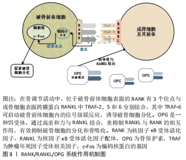
[57] CHEN B, WU W, SUN W, et al. RANKL expression in periodontal disease: where does RANKL come from? Biomed Res Int. 2014;2014:731039.
[58] BARBATO L, FRANCIONI E, BIANCHI M, et al. Periodontitis and bone metabolism. Clin Cases Miner Bone Metab. 2015;12(2):174-177.
[59] 李小娜.OPG/RANK/RANKL信号通路研究进展[J].河南医学研究, 2018,27(10):1802-1804.
[60] Sojod B, Chateau D, Mueller CG, et al. RANK/RANKL/OPG signalization implication in periodontitis: new evidence from a RANK transgenic mouse model. Front Physiol. 2017;8:338.
[61] 邹林洪,胡丹,张琳林,等.OPG/RANKL/RANK调节系统与大鼠慢性牙周炎发展的相关性分析[J].重庆医学,2016,45(31):4334-4336.
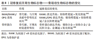
[62] 林志明,陈宏柏,彭秀燕.两种冠修复材料对牙周状况及龈沟液中TWEAK、RANKL水平的影响[J].临床口腔医学杂志,2019,35(8): 467-470.
[63] 章洁,徐国超.骨桥蛋白在牙周炎防治中的影响研究[J].中国卫生检验杂志,2017,27(4):511-512,515.
|