中国组织工程研究 ›› 2022, Vol. 26 ›› Issue (5): 700-705.doi: 10.12307/2022.114

• 组织构建实验造模 experimental modeling in tissue construction • 上一篇    下一篇

金黄色葡萄球菌生物膜克氏针置入建立创伤性大鼠骨髓炎模型

冯建波1,2,李陈诚1,2,刘金月1,2,王小敏3,彭笳宸1,2   

  1. 1遵义医科大学附属医院关节外科,贵州省遵义市   563000;2遵义医科大学-罗切斯特大学联合骨科研究中心,贵州省遵义市   563000;3遵义医科大学,贵州省遵义市   563000 
  • 收稿日期:2020-10-29 修回日期:2020-10-30 接受日期:2020-12-07 出版日期:2022-02-18 发布日期:2021-10-28
  • 通讯作者: 彭笳宸,博士,主任医师/教授,遵义医科大学附属医院关节外科,贵州省遵义市 563000;遵义医科大学-罗切斯特大学联合骨科研究中心,贵州省遵义市 563000 并列通讯作者:王小敏,博士,副教授,遵义医科大学基础医学院微生物实验室,贵州省遵义市 563000
  • 作者简介:冯建波,男,1992年生,贵州省遵义市人,2015年遵义医科大学毕业,医师,主要从事骨关节感染研究。
  • 基金资助:
    贵州省科技计划项目(黔科合支撑[2018]2760),项目负责人:彭笳宸;国家自然科学基金项目(81760400),项目负责人:彭笳宸

Implantation of Kirschner wire with Staphylococcus aureus biofilm establishes a traumatic osteomyelitis model in rats

Feng Jianbo1, 2, Li Chencheng1, 2, Liu Jinyue1, 2, Wang Xiaomin3, Peng Jiachen1, 2   

  1. 1Department of Joint Surgery, Affiliated Hospital of Zunyi Medical University, Zunyi 563000, Guizhou Province, China; 2Zunyi Medical University-University of Rochester Joint Orthopedic Research Center, Zunyi 563000, Guizhou Province, China; 3Zunyi Medical University, Zunyi 563000, Guizhou Province, China 
  • Received:2020-10-29 Revised:2020-10-30 Accepted:2020-12-07 Online:2022-02-18 Published:2021-10-28
  • Contact: Peng Jiachen, MD, Chief physician, Professor, Department of Joint Surgery, Affiliated Hospital of Zunyi Medical University, Zunyi 563000, Guizhou Province, China; Zunyi Medical University-University of Rochester Joint Orthopedic Research Center, Zunyi 563000, Guizhou Province, China Co-corresponding author: Wang Xiaomin, MD, Associate professor, Zunyi Medical University, Zunyi 563000, Guizhou Province, China
  • About author:Feng Jianbo, Physician, Department of Joint Surgery, Affiliated Hospital of Zunyi Medical University, Zunyi 563000, Guizhou Province, China; Zunyi Medical University-University of Rochester Joint Orthopedic Research Center, Zunyi 563000, Guizhou Province, China
  • Supported by:
    The Science and Technology Plan Project of Guizhou Province, No. [2018]2760 (to PJC); National Natural Science Foundation of China, No. 81760400 (to PJC) 

摘要:

文题释义:
骨髓炎:为一种骨的感染和破坏,可由需氧或厌氧菌、分枝杆菌及真菌引起,骨髓炎好发于长骨、糖尿病患者的足部或由于创伤或手术引起的穿透性骨损伤部位。
生物膜:也称为生物被膜,是指附着于有生命或无生命物体表面被细菌胞外大分子包裹的有组织的细菌群体,生物膜细菌对抗生素和宿主免疫防御机制的抗性很强。

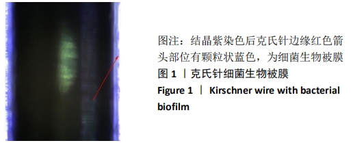
背景:建立可靠且接近临床的动物模型是研究骨髓炎治疗方法的基础和关键,以往均为直接滴加细菌至钻孔内制作骨髓炎模型,与临床发病因素有明显区别,且滴入细菌悬液量不易控制,易导致模型动物死亡,而在临床中以细菌生物膜导致骨髓炎的情况最多见。
目的:利用带细菌生物膜克氏针置入骨髓腔来制作大鼠骨髓炎模型。
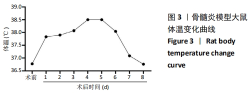
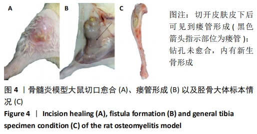
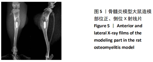
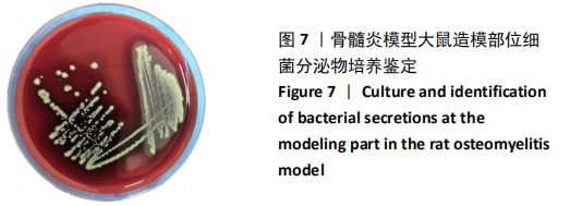
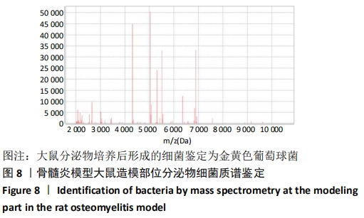
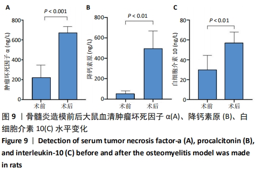
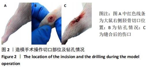
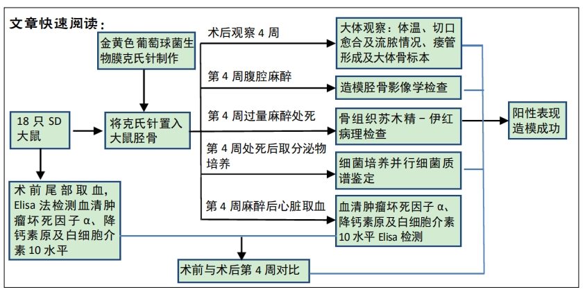
方法:用金黄色葡萄球菌制作带细菌生物膜克氏针,取18只健康SD大鼠,将带金黄色葡萄球菌生物膜克氏针置入SD大鼠胫骨,以无菌石蜡封闭钻孔。造模后密切观察动物行动状态及切口愈合情况,4周后行影像学及病理学检查观察骨感染情况,无菌取造模部位分泌物作细菌培养,并行质谱鉴定其是否为制作细菌生物膜时接种的细菌,并检测造模前后大鼠血清炎症因子,作为评估骨髓炎模型造模成功的指标。
结果与结论:18只大鼠均符合骨髓炎特征,均表现出不同程度的流脓、死骨、死腔以及新生骨形成;造模后大鼠降钙素原、肿瘤坏死因子α以及白细胞介素10等血清炎症因子增高明显(P < 0.05);经过鉴定大鼠分泌物培养后形成的细菌为金黄色葡萄球菌。以上结果证实:通过金黄色葡萄球菌生物膜克氏针置入大鼠胫骨可以成功制作大鼠骨髓炎模型,且更接近临床骨髓炎发病情况。 

https://orcid.org/0000-0003-2126-1325 (冯建波);https://orcid.org/0000-0001-7267-0650 (彭笳宸) 

关键词: 骨髓炎, 大鼠, 动物模型, 金黄色葡萄球菌, 细菌生物膜, 克氏针

Abstract:

BACKGROUND: To establish an animal osteomyelitis model is the basis and key to explore the treatment of osteomyelitis. In the past, direct addition of bacteria was used to make the osteomyelitis model, which is significantly different from clinical pathogenic factors. Moreover, it is difficult to control the amount of bacterial suspension dripped, which can easily lead to the death of model animals. Osteomyelitis caused by bacterial biofilms is the most common in clinical practice. 
OBJECTIVE: To prepare a rat model of osteomyelitis by inserting Kirschner wires with bacterial biofilms into the rat bone marrow cavity.
METHOD: Bacterium-bearing Kirschner wires were prepared using Staphylococcus aureus. Eighteen healthy Sprague-Dawley rats were enrolled. Kirschner wires with Staphylococcus aureus biofilms were inserted into the rat tibia, and the borehole was sealed with sterile paraffin. Rats’ behaviors and incision healing were observed after modeling. Bone infection was observed through imaging and pathological examinations at 4 weeks after modeling. Secretions at the model site were aseptically taken for bacterial culture, and mass spectrometry was used to identify whether they were the bacteria inoculated during the production of bacterial biofilms, and the serum inflammatory factors of rats before and after modeling were detected as indicators for evaluating the success of the osteomyelitis model.
RESULTS AND CONCLUSION: All the 18 rats presented with the characteristics of osteomyelitis. They all showed different degrees of pus, dead bone, dead space and new bone formation. After modeling, the levels of serum inflammatory factors, such as procalcitonin, tumor necrosis factor alpha and interleukin-10, increased significantly (P < 0.05). The bacteria that formed after secretion culture were identified as Staphylococcus aureus. To conclude, the rat osteomyelitis model can be successfully made by inserting the Kirschner wire into the rat tibia through the Staphylococcus aureus biofilm, which is closer to osteomyelitis in clinic.

Key words: osteomyelitis, rats, animal model, Staphylococcus aureus, bacterial biofilm, Kirschner wire

中图分类号: