中国组织工程研究 ›› 2022, Vol. 26 ›› Issue (2): 225-231.doi: 10.12307/2022.037

• 组织构建实验造模 experimental modeling in tissue construction • 上一篇    下一篇

电针干预大鼠大脑中动脉栓塞缺血再灌注模型丝裂原活化蛋白激酶通路的变化

赖  涵1,王  娇1,董苗苗1,罗  梦1,王文豪1,周国平1,2   

  1. 1南方医科大学中西医结合医院,广东省广州市   510315;2南方医科大学中医药学院,广东省广州市   510515
  • 收稿日期:2020-09-17 修回日期:2020-09-17 接受日期:2020-10-30 出版日期:2022-01-18 发布日期:2021-10-27
  • 通讯作者: 周国平,主任医师,博士生导师,南方医科大学中西医结合医院,广东省广州市 510315;南方医科大学中医药学院,广东省广州市 510515
  • 作者简介:赖涵,女,1994年生,南方医科大学在读硕士,主要从事中风病的机制研究。
  • 基金资助:
    国家自然科学基金(81674048),项目负责人:周国平

Electroacupuncture intervenes with changes of mitogen-activated protein kinase pathway in a rat model of cerebral ischemia/reperfusion due to middle cerebral artery occlusion

Lai Han1, Wang Jiao1, Dong Miaomiao1, Luo Meng1, Wang Wenhao1, Zhou Guoping1, 2   

  1. 1Hospital of Integrated Traditional Chinese Medicine and Western Medicine, Southern Medical University, Guangzhou 510315, Guangdong Province, China; 2School of Traditional Chinese Medicine, Southern Medical University, Guangzhou 510515, Guangdong Province, China
  • Received:2020-09-17 Revised:2020-09-17 Accepted:2020-10-30 Online:2022-01-18 Published:2021-10-27
  • Contact: Zhou Guoping, Chief physician, Doctoral supervisor, Hospital of Integrated Traditional Chinese Medicine and Western Medicine, Southern Medical University, Guangzhou 510315, Guangdong Province, China; School of Traditional Chinese Medicine, Southern Medical University, Guangzhou 510515, Guangdong province, China
  • About author:Lai Han, Master candidate, Hospital of Integrated Tranditonal Chinese Medicine and Western Medicine, Southern Medical University, Guangzhou 510315, Guangdong Province, China
  • Supported by:
    the National Natural Science Foundation of China, No. 81674048 (to ZGP)

摘要:

文题释义:
丝裂原活化蛋白激酶(mitogen acti-vatedprotrinkinase,MAPK)家族:MAPK在神经细胞的生长和增殖中起作用,MAPK信号通路在神经系统中广泛表达,并调节脑缺血再灌注损伤后神经细胞的修复或凋亡。MAPK的3个主要亚家族;细胞外信号调节激酶(ERK)、c-Jun N端激酶(JNK)和p38激酶参与调节脑缺血再灌注损伤后神经元细胞的功能障碍。
脑缺血再灌注损伤:脑缺血再灌注会导致炎症、氧化损伤和程序性细胞死亡,从而加剧脑组织损伤并严重影响中风患者的日常生活,当脑缺血再灌注发生时,MAPK家族开始发生调控作用。
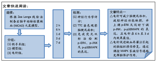
背景既往电针对大鼠脑缺血再灌注损伤保护作用及时效性的机制研究较少。
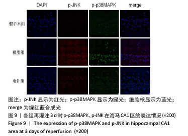
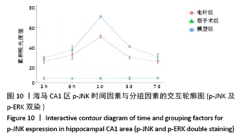
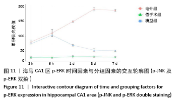
目的:观察不同时间电针对脑缺血再灌注大鼠MAPK家族3条信号通路中p-ERK、p-JNK及p-p38MAPK蛋白表达的影响,探索电针发挥脑保护作用的具体机制。
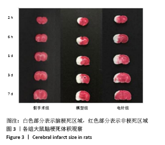
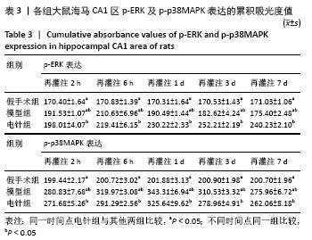
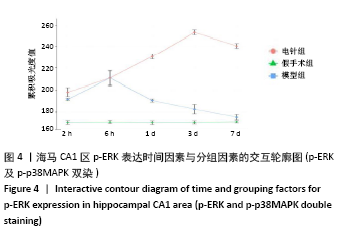
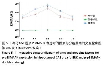
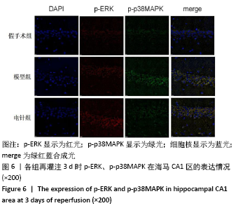
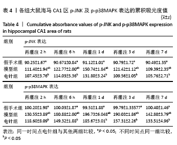
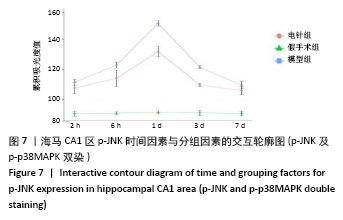
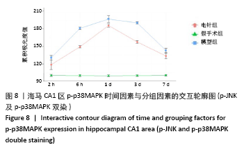
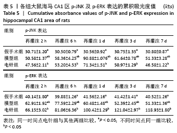
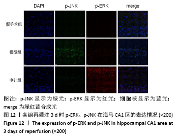
方法:150只SD雄性大鼠随机分为假手术组、模型组、电针组(后2组线栓法制备右侧大脑中动脉栓塞缺血模型),每组50只,每组再设脑缺血1.5 h后再灌注2 h、6 h、1 d、3 d、7 d共5个时间亚组,每亚组10只。电针各亚组在规定时间点行单次电针治疗,选取“合谷”“尺泽”“足三里”“三阴交”,予疏密波,频率2 Hz,强度1 mA,治疗20 min;电针的同一时间抓取模型组和假手术组的大鼠进行固定,但不进行电针干预。在相应时间点进行神经功能缺损评分,之后大鼠麻醉后取脑,TTC染色法观察脑梗死面积,免疫荧光双标法结合检测p-ERK、p-JNK、p-p38MAPK的表达。
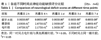
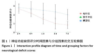
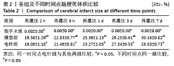
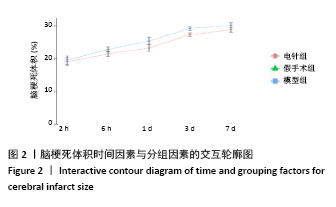
结果与结论:①与假手术组相比,模型组同时段各亚组神经功能缺损评分升高、脑梗死体积增大(P < 0.05);与模型组相比,电针组再灌注6 h、1 d、3 d亚组神经功能缺损评分显著降低、脑梗死体积明显减少(P < 0.05);②与模型组比较,电针组再灌注1,3 d出现p-ERK表达的明显上调(P < 0.05)及p-JNK的表达显著下降(P < 0.05),p-p38MAPK在再灌注6 h、3 d时下降明显再灌注;③结果说明电针可减少脑梗死体积,减轻神经功能缺损,并上调p-ERK同时下调p-JNK、p-p38MAPK的表达,且电针在再灌注6 h-3 d内效果最佳;电针促进缺血再灌注引起的脑组织损伤修复,通过调控MAPK家族信号通路发挥脑保护作用。

https://orcid.org/0000-0002-4613-4384 (周国平) 

中国组织工程研究杂志出版内容重点:组织构建;骨细胞;软骨细胞;细胞培养;成纤维细胞;血管内皮细胞;骨质疏松;组织工程

关键词: 电针, 脑缺血再灌注, MAPK通路, 脑梗死, 大鼠

Abstract: BACKGROUND: The mechanisms underlying the protection of electroacupuncture against cerebral ischemia/reperfusion injury in rats and its temporal efficacy have been less studied in the past.
OBJECTIVE: To observe the effect of electroacupuncture on the expression of p-ERK, p-JNK and p-p38MAPK proteins in three signaling pathways of the MAPK family in rats with cerebral ischemia/reperfusion at different times, and to explore the specific mechanism underlying the cerebral protection of electroacupuncture.
METHODS: 150 Sprague-Dawley male rats were randomly divided into sham-operated group, model group, and electroacupuncture group, with 50 rats in each group. Each group was then set up for ischemia 1.5 hours followed by reperfusion for 2 hours, 6 hours, 1 day, 3 days, and 7 days, with 10 rats in each subgroup. Single electroacupuncture treatment at Hegu, Shakuze, Zusanli, and Sanyinjiao was conducted at the given time, giving sparse waves at a frequency of 2 Hz, intensity of 1 mA, 20 minutes for one session. Rats in the model and sham-operated groups were grasped simultaneously for fixation at the same time of electroacupuncture, but no electroacupuncture intervention was performed. After anesthesia, the brains were decapitated at the corresponding time points, and neurological deficit scoring was used to observe the infarct area by TTC staining. The expression of p-ERK, p-JNK, and p-p38MAPK was detected by double immunofluorescence labeling method.
RESULTS AND CONCLUSION: Compared with the sham-operated group, the model group had higher neurological deficit scores and larger infarct size in each subgroup in the same period (P < 0.05). Compared with the model group, there were significantly lower neurobehavioral scores and smaller infarct size in the electroacupuncture subgroups of 6 hours, 1 day and 3 days (P < 0.05). Compared with the model group, the electroacupuncture group had significant up-regulation of p-ERK expression at 1 and 3 days of reperfusion (P < 0.05), downregulation of p-JNK expression at 1 and 3 days of reperfusion (P < 0.05), and significant downregulation of p-p38MAPK expression at 6 hours and 3 days of reperfusion (P < 0.05). To conclude, electroacupuncture can reduce infarct size, alleviate neurological deficits, up-regulate p-ERK expression and down-regulate the expression of p-JNK and p-p38MAPK at the same time. The best effect of electroacupuncture is within 6 hours-3 days. Electroacupuncture promotes the repair of brain tissue damage caused by cerebral ischemia/reperfusion and exerts cerebral protection through the regulation of MAPK family signaling pathway.


Key words: electroacupuncture, cerebral ischemia/reperfusion, MAPK signaling pathway, cerebral infarction, rat

中图分类号: