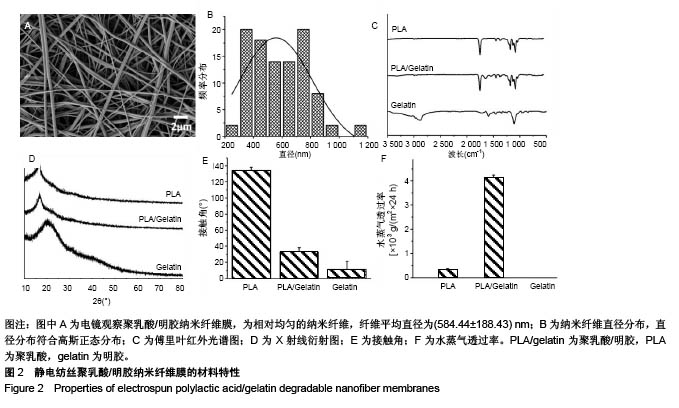
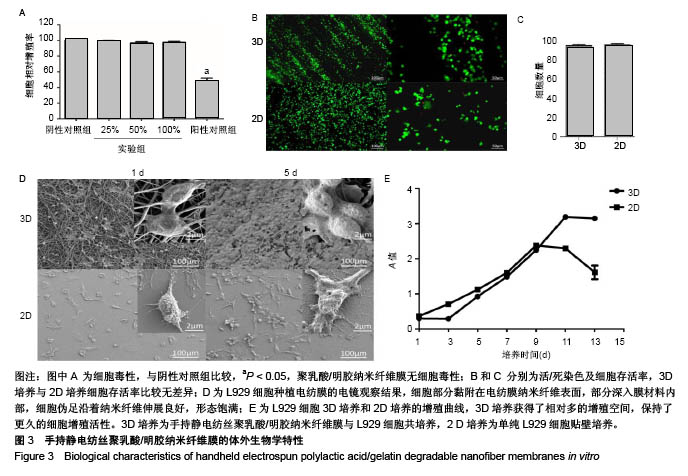
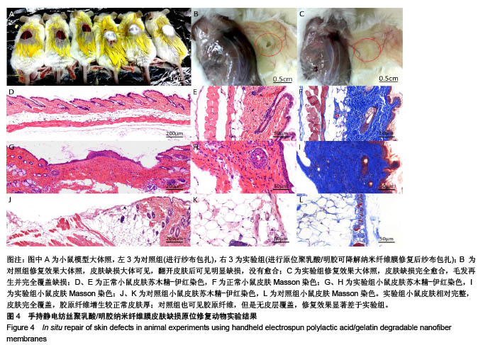
| [1] Pérez-Sánchez A, Barrajón-Catalán E, Herranz-López M, et al. Nutraceuticals for Skin Care: A Comprehensive Review of Human Clinical Studies. Nutrients. 2018;10(4). pii: E403. doi: 10. 3390/nu10040403. [2] Sanford JA, Gallo RL. Functions of the skin microbiota in health and disease. Semin Immunol. 2013;25(5):370-377. [3] Swann G. The skin is the body's largest organ. J Vis Commun Med. 2010;33(4):148-149. [4] Na M, Mohammad M, Fei Y, et al. Lack of Receptor for Advanced Glycation End products leads to less severe staphylococcal skin infection but more skin abscesses and prolonged wound healing. J Infect Dis. 2018. doi:10. 1093/infdis/jiy007. [Epub ahead of print][5] Albright V, Xu M, Palanisamy A, et al. Micelle-Coated, Hierarchically Structured Nanofibers with Dual-Release Capability for Accelerated Wound Healing and Infection Control. Adv Healthc Mater. 2018:e1800132. [6] Sadiq A, Shah A, Jeschke MG, et al. The Role of Serotonin during Skin Healing in Post-Thermal Injury. Int J Mol Sci. 2018;19(4). pii: E1034. [7] Shi L, Zhao Y, Xie Q, et al. Moldable Hyaluronan Hydrogel Enabled by Dynamic Metal-Bisphosphonate Coordination Chemistry for Wound Healing. Adv Healthc Mater. 2018;7(5). doi:10. 1002/adhm. 201700973. [8] Shin YC, Shin DM, Lee EJ, et al. Hyaluronic Acid/PLGA Core/Shell Fiber Matrices Loaded with EGCG Beneficial to Diabetic Wound Healing. Adv Healthc Mater. 2016;5(23): 3035-3045. [9] Wang S, Yang H, Tang Z, et al. Wound Dressing Model of Human Umbilical Cord Mesenchymal Stem Cells-Alginates Complex Promotes Skin Wound Healing by Paracrine Signaling. Stem Cells Int. 2016;2016:3269267. [10] Hsu LC, Peng BY, Chen MS, et al. The potential of the stem cells composite hydrogel wound dressings for promoting wound healing and skin regeneration: In vitro and in vivo evaluation. J Biomed Mater Res B Appl Biomater. 2018. doi:10. 1002/jbm. b. 34118. [Epub ahead of print][11] Zarei F, Soleimaninejad M. Role of growth factors and biomaterials in wound healing. Artif Cells Nanomed Biotechnol. 2018;15:1-6. [12] Sheikholeslam M, Wright MEE, Jeschke MG, et al. Biomaterials for Skin Substitutes. Adv Healthc Mater. 2018;7(5). doi: 10.1002/adhm.201700897.[13] Zhang W, Chen L, Chen J, et al. Silk Fibroin Biomaterial Shows Safe and Effective Wound Healing in Animal Models and a Randomized Controlled Clinical Trial. Adv Healthc Mater. 2017;6(10). doi:10.1002/adhm. 201700121. [14] Paschoalin RT, Traldi B, Aydin G, et al. Solution blow spinning fibres: New immunologically inert substrates for the analysis of cell adhesion and motility. Acta Biomater. 2017;51:161-174. [15] Su K, Wang C. Recent advances in the use of gelatin in biomedical research. Biotechnol Lett. 2015;37(11):2139-2145. [16] Deng K, Yang Y, Ke Y, et al. A novel biomimetic composite substitute of PLLA/gelatin nanofiber membrane for dura repairing. Neurol Res. 2017;39(9):819-829. [17] Liu Y, Cui H, Zhuang X, et al. Electrospinning of aniline pentamer-graft-gelatin/PLLA nanofibers for bone tissue engineering. Acta Biomater. 2014;10(12):5074-5080. [18] 陈亮,许运.静电纺丝微米/纳米纤维材料在硬膜组织损伤修复中的应用[J].中华实验外科杂志,2016,33(7):1880-1883.[19] Xu SC, Qin CC, Yu M, et al. A battery-operated portable handheld electrospinning apparatus. Nanoscale. 2015;7(29): 12351-12355. [20] Dong RH, Jia YX, Qin CC, et al. In situ deposition of a personalized nanofibrous dressing via a handy electrospinning device for skin wound care. Nanoscale. 2016;8(6):3482-3488. [21] Dong RH, Qin CC, Qiu X, et al. In situ precision electrospinning as an effective delivery technique for cyanoacrylate medical glue with high efficiency and low toxicity. Nanoscale. 2015;7(46):19468-19475. [22] Lv FY, Dong RH, Li ZJ, et al. In situ precise electrospinning of medical glue fibers as nonsuture dural repair with high sealing capability and flexibility. Int J Nanomedicine. 2016;11: 4213-4220. [23] Deng K, Ye X, Yang Y, et al. Evaluation of efficacy and biocompatibility of a new absorbable synthetic substitute as a dural onlay graft in a large animal model. Neurol Res. 2016; 38(9):799-808. [24] van Wachem PB, Hogt AH, Beugeling T, et al. Adhesion of cultured human endothelial cells onto methacrylate polymers with varying surface wettability and charge. Biomaterials. 1987;8(5):323-328. [25] Kailani MH, Jafar H, Awidi A. Synthetic Biomaterials for Skin Tissue Engineering[M]// Skin Tissue Engineering and Regenerative Medicine, 2016:163-183. [26] 郑崇亮,陆树良.创伤后表皮更新和修复的研究进展[J].中华创伤杂志,2017,33(2):185-188.[27] 杨道朋,夏旭.3D打印生物材料研究及其临床应用优势[J].中国组织工程研究,2017,22(18):2927-2933.[28] Dash BC, Xu Z, Lin L, et al. Stem Cells and Engineered Scaffolds for Regenerative Wound Healing. Bioengineering (Basel). 2018;5(1). pii: E23. doi: 10. 3390/bioengineering 5010023. Review. [29] Sun L, Gao W, Fu X, et al. Enhanced wound healing in diabetic rats by nanofibrous scaffolds mimicking the basketweave pattern of collagen fibrils in native skin. Biomater Sci. 2018;6(2):340-349. [30] Lien SM, Ko LY, Huang TJ. Effect of pore size on ECM secretion and cell growth in gelatin scaffold for articular cartilage tissue engineering. Acta Biomater. 2009;5(2): 670-679. [31] Hoveizi E, Ebrahimi-Barough S, Tavakol S, et al. In Vitro Differentiation of Human iPS Cells into Neural like Cells on a Biomimetic Polyurea. Mol Neurobiol. 2017;54(1):601-607. [32] Kim HW, Yu HS, Lee HH. Nanofibrous matrices of poly(lactic acid) and gelatin polymeric blends for the improvement of cellular responses. J Biomed Mater Res A. 2008;87(1):25-32. [33] Wang S, Zhang Y, Wang H, et al. Fabrication and properties of the electrospun polylactide/silk fibroin-gelatin composite tubular scaffold. Biomacromolecules. 2009;10(8):2240-2244. [34] Chiou BS, Jafri H, Avena-Bustillos R, et al. Properties of electrospun pollock gelatin/poly(vinyl alcohol) and pollock gelatin/poly(lactic acid) fibers. Int J Biol Macromol. 2013; 55:214-220. [35] Shi Z, Xu T, Yuan Y, et al. A New Absorbable Synthetic Substitute With Biomimetic Design for Dural Tissue Repair. Artif Organs. 2016;40(4):403-413. [36] Haik J, Kornhaber R, Blal B, et al. The Feasibility of a Handheld Electrospinning Device for the Application of Nanofibrous Wound Dressings. Adv Wound Care(New Rochelle). 2017;6(5):166-174. [37] Sofokleous P, Stride E, Bonfield W, et al. Design, construction and performance of a portable handheld electrohydrodynamic multi-needle spray gun for biomedical applications. Mater Sci Eng C Mater Biol Appl. 2013;33(1):213-223. [38] 林青,郝宏,王坤,等.逆向设计在制造工程中的应用[J].新技术新工艺,2017,37(12):65-67.[39] Wong JY, Pfahnl AC. 3D Printed Surgical Instruments Evaluated by a Simulated Crew of a Mars Mission. Aerosp Med Hum Perform. 2016;87(9):806-810. [40] Azimifar F, Hassani K, Saveh AH, et al. A medium invasiveness multi-level patient's specific template for pedicle screw placement in the scoliosis surgery. Biomed Eng Online. 2017;16(1):130. |
.jpg)
.jpg)
.jpg)