中国组织工程研究 ›› 2014, Vol. 18 ›› Issue (28): 4463-4468.doi: 10.3969/j.issn.2095-4344.2014.28.006

• 骨髓干细胞 bone marrow stem cells • 上一篇    下一篇

密度梯度离心及贴壁分离筛选相结合分离培养大鼠骨髓间充质干细胞

王英慧,郑瑞,陈莉   

  1. 武警后勤学院临床医学系实验技术教研室,天津市  300162
  • 出版日期:2014-07-02 发布日期:2014-07-02
  • 作者简介:王英慧,女,1974年生,河北省丰润县人,汉族,2012年河北医科大学毕业,硕士,实验师,主要从事细胞分化的研究。

Isolation and culture of rat bone marrow mesenchymal stem cells using density gradient centrifugation and adherence separation screening

Wang Ying-hui, Zheng Rui, Chen Li   

  1. Research Room of Experimental Technology, Department of Clinical Medicine, Logistics University of People’s Armed Police Force, Tianjin 300162, China
  • Online:2014-07-02 Published:2014-07-02
  • About author:Wang Ying-hui, Master, Experimentalist, Research Room of Experimental Technology, Department of Clinical Medicine, Logistics University of People’s Armed Police Force, Tianjin 300162, China

摘要:

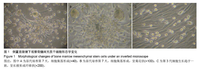
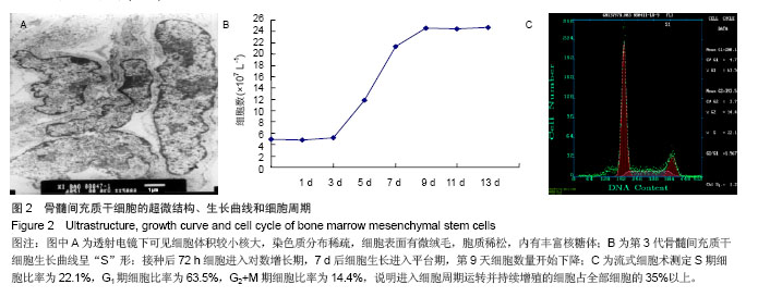
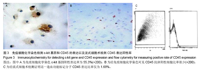
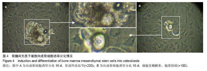
背景:在正常情况下骨髓来源间充质干细胞含量很少,且易与其他细胞相混杂,因此,建立一种简便可行的体外培养扩增方法,获得大量稳定的骨髓间充质干细胞具有重要的理论意义和应用价值。
目的:建立一种简便可行的体外培养扩增方法,获得大量稳定的骨髓间充质干细胞。
方法:采用密度梯度离心法及贴壁分离筛选法相结合体外分离培养SD大鼠骨髓间充质干细胞。倒置光学显微镜下观察细胞形态变化,透射电镜观察细胞亚微结构,锥虫蓝拒染法计算活细胞数,绘制细胞生长曲线,流式细胞仪分析细胞周期,免疫细胞化学检测c-kit和CD45的表达,流式细胞仪分析CD45的表达情况。
结果与结论: ①接种后24 h倒置光学显微镜下可见有细胞贴壁并伸出伪足,4 d时可见有细胞集落形成,14 d时细胞可达到90%融合。经传代后细胞趋于一致,为纤维样细胞,呈漩涡或火焰状生长。②透射电镜下可见第3代骨髓间充质干细胞体积较小核大,核仁明显。染色质分布稀疏,电子密度低。细胞表面有微绒毛,胞质稀松,内有丰富核糖体,而内质网、线粒体、高尔基复合体等细胞器少见,提示细胞处于原始未分化状态。③第3代骨髓间充质干细胞接种后第1天细胞数量有所减少,第2天细胞开始增长,第3天细胞进入指数增生期,第7天进入平台期,第9天细胞数量开始下降,绘制的生长曲线呈“S”形。④第3代骨髓间充质干细胞经DNA染色后采用流式细胞仪测定其DNA含量证明S期细胞比率为21.1%。⑤免疫细胞化学染色和流式细胞术证明c-kit阳性细胞比率为53.3%,CD45表达阳性率为1.68%。⑥骨髓间充质干细胞向成骨细胞诱导分化16 d后,细胞的胞体呈椭圆形,有短突起彼此相连,胞浆较暗,提示可能富含粗面内质网和高尔基复合体,并具有分泌类骨质的功能。结果证明该方法获得的大鼠骨髓间充质干细胞生长稳定,增殖活跃。


中国组织工程研究杂志出版内容重点:干细胞;骨髓干细胞;造血干细胞;脂肪干细胞;肿瘤干细胞;胚胎干细胞;脐带脐血干细胞;干细胞诱导;干细胞分化;组织工程


全文链接:

关键词: 干细胞, 骨髓干细胞, 贴壁分离筛选法, 骨髓间充质干细胞, 生物学特性, SD大鼠, 细胞形态, 细胞周期, 增殖

Abstract:

BACKGROUND: Bone marrow mesenchymal stem cell content is less under normal conditions, and easily confounded with other cells. Therefore, to establish a simple feasible in vitro cultured amplification method and to obtain a large number of stable bone marrow mesenchymal stem cells are of important theoretical significance and application value.
OBJECTIVE: To establish a simple feasible in vitro culture and amplification method and to obtain numerous stable bone marrow mesenchymal stem cells.
METHODS: Bone marrow mesenchymal stem cells were isolated from Sprague-Dawley rats and cultured in vitro using density gradient centrifugation and adherence separation screening. Cell morphological changes were observed under an inverted light microscope. Submicroscopic structure of cells was observed under a transmission electron microscope. Living cell number was counted using Trypan blue exclusion method. Cell growth curve was drawn. Cell cycle was analyzed using flow cytometry. The expression of c-kit and CD45 was measured using immunocytochemistry. CD45 expression was analyzed using flow cytometry.
RESULTS AND CONCLUSION: (1) Cell adhesion and pseudopodia could be seen under an inverted light microscope at 24 hours after inoculation; cell colony formed at 4 days; 90% cell confluence was found at 14 days. After passage, the cells were tended to homogenous and became fibroblast-like cells, which showed whirl-like growing or flamboyancy growing. (2) The size of passage 3 bone marrow mesenchymal stem cells was small and nucleolus was large, with clear nucleoli under a transmission electron microscope, with the presence of sparse chromatin and low electron density. There was microvillus on the surface of cells, abundant ribosomes could be seen in the cytoplasm with few other cellular organs such as endoplasmic reticulum, mitochondria and Golgi complex. These showed that ultrastructural structure had undifferentiated features. (3) The number of passage 3 bone marrow mesenchymal stem cells was reduced at 1 day after inoculation. The cells began to grow at 2 days, and entered the period of exponential growth at 3 days, entered the flat period at 7 days and the number of cells began to decrease at 9 days. Growth curve exhibited “S” shape. (4) The percentage of S phase cells was 21.1% as detected by flow cytometry after passage 3 bone marrow mesenchymal stem cells were stained by DNA. (5) Immunocytochemistry and flow cytometry had proved that the c-kit positive rate of cells was 53.3% and CD45 positive rate was 1.68%. (6) After osteogenic induction of mesenchymal stem cells for 16 days, cells became oval, and short processes connected each other. The cytoplasm was dark, suggesting that it may be rich in rough endoplasmic reticulum and Golgi complex. They can secrete osteoid. Results verified that obtained bone marrow mesenchymal stem cells stably grew, and actively proliferated.


中国组织工程研究杂志出版内容重点:干细胞;骨髓干细胞;造血干细胞;脂肪干细胞;肿瘤干细胞;胚胎干细胞;脐带脐血干细胞;干细胞诱导;干细胞分化;组织工程


全文链接:

Key words: bone marrow, mesenchymal stem cells, cells, cultured, rats, Sprague-Dawley

中图分类号: